. 2020 May;57(5):2232-2243.
doi: 10.1007/s12035-020-01868-8. Epub 2020 Jan 29.

Selected microRNAs Increase Synaptic Resilience to the Damaging Binding of the Alzheimer's Disease Amyloid Beta Oligomers

Affiliations

Selected microRNAs Increase Synaptic Resilience to the Damaging Binding of the Alzheimer's Disease Amyloid Beta Oligomers

Olga Zolochevska et al. Mol Neurobiol. 2020 May.

Abstract

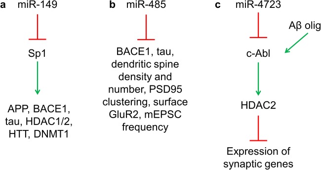
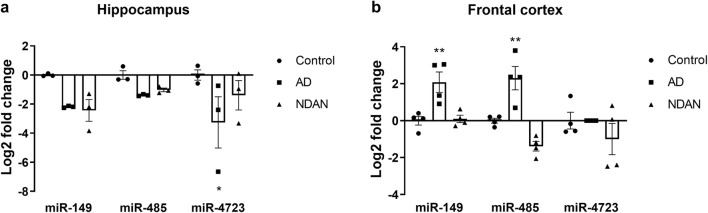
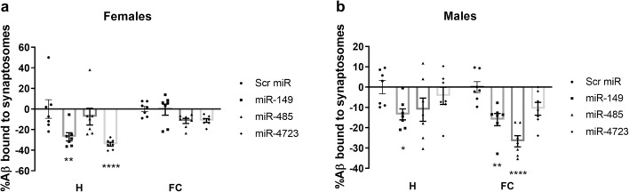
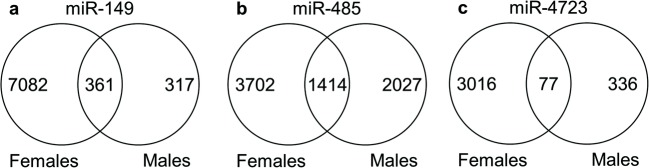
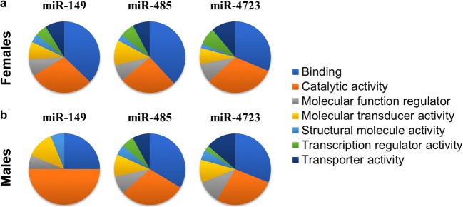
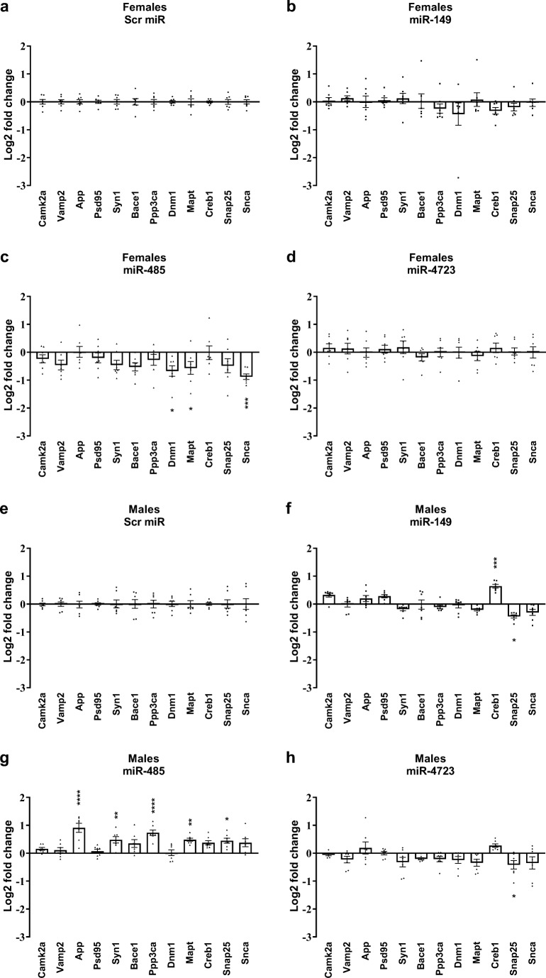
Alzheimer's disease (AD) is marked by synaptic loss (at early stages) and neuronal death (at late stages). Amyloid beta (Aβ) and tau oligomers can target and disrupt synapses thus driving cognitive decay. Non-demented individuals with Alzheimer's neuropathology (NDAN) are capable of withstanding Aβ and tau toxicity, thus remaining cognitively intact despite presence of AD neuropathology. Understanding the involved mechanism(s) would lead to development of novel effective therapeutic strategies aimed at promoting synaptic resilience to amyloid toxicity. NDAN have a unique hippocampal post-synaptic proteome when compared with AD and control individuals. Potential upstream modulators of such unique proteomic profile are miRNA-485, miRNA-4723 and miRNA-149, which we found differentially expressed in AD and NDAN vs. control. We thus hypothesized that these miRNAs play an important role in promoting either synaptic resistance or sensitization to Aβ oligomer binding. Using an in vivo mouse model, we found that administration of these miRNAs affected key synaptic genes and significantly decreased Aβ binding to the synapses. Our findings suggest that miRNA regulation and homeostasis are crucial for Aβ interaction with synaptic terminals and support that a unique miRNA regulation could be driving synaptic resistance to Aβ toxicity in NDAN, thus contributing to their preserved cognitive abilities.

Keywords: Alzheimer’s disease; Non-demented with Alzheimer’s neuropathology; Synaptic resilience; microRNA.


Figures

Fig. 1
Fig. 1
Functions of miR-149, miR-485, and miR-4723. miR-149 (a), miR-485 (b), and miR-4723 (c) are involved in regulation of synaptic genes. Sp1 specificity protein 1, APP amyloid precursor protein, BACE1 beta-secretase 1, HDAC1/2 histone deacetylase 1/2, HTT huntingtin, DNMT1 DNA methyltransferase, PSD95 post-synaptic density protein 95, GluR2 glutamate receptor 2, mEPSC miniature excitatory post-synaptic currents, c-Abl Abelson tyrosine-protein kinase 1. References are provided in text
Fig. 2
Fig. 2
miR levels in the hippocampus of control, AD, and NDAN. IPA-predicted miR-149, miR-485, and miR-4723 are differentially regulated in post-mortem hippocampi (a) and frontal cortices (b) of AD and NDAN when compared with control, which is set at zero. miR-4723 was below detection limit in AD frontal cortex. Measured miR values were normalized to the expression level of U6. Values represent the means ± SEM. n = 4 frontal cortex, n = 3 hippocampus. *p < 0.05 and **p < 0.01 vs. control, two-way ANOVA, followed by Dunnett’s multiple comparisons test
Fig. 3
Fig. 3
Aβ oligomer binding to synaptosomes in mice after ICV treatment with miR-149, miR-485, and miR-4723. Female (a) and male (b) mice were injected ICV with the selected miRs; scrambled miR was used as control. Synaptosomes were isolated from hippocampi (H) and frontal cortices (FC) and incubated ex vivo with 2.5 μM tagged Aβ oligomers and analyzed using flow cytometry. Levels of binding to scrambled-injected mice were set at zero. n = 7. Values represent the means ± SEM. *p < 0.05, **p < 0.01, and ****p < 0.0001 vs. scrambled miR, two-way ANOVA, followed by Dunnett’s multiple comparisons test.
Fig. 4
Fig. 4
Changes in the hippocampal transcriptome after treatment with miR-149, miR-485, and miR-4723. RNA changes in the hippocampi of miR-treated animals were normalized to mice injected with scrambled miR. The Venn diagram shows an overlap in RNA changes in males vs. females. n = 3 mice/group. The Venn diagrams were built using the online Venny tool [69]
Fig. 5
Fig. 5
Hippocampal transcriptome analyzed with PANTHER. PANTHER [70, 71] was used to analyze the molecular functions of mRNAs changed after a female and b male mice were treated ICV with miR-149, miR-485, and miR-4723. Changes in miR-treated animals were normalized to mice injected with scrambled miR
Fig. 6
Fig. 6
Expression of synaptic genes in hippocampi after treatment with miR-149, miR-485, and miR-4723. Several genes involved in synaptic function were assessed using qRT-PCR in hippocampi obtained from ad female or eh male mice treated with miRs. Mice treated with scrambled miR were used as a control and set at zero. Measured mRNA levels were normalized to the expression level of actin. Values represent the means ± SEM. n = 7. *p < 0.05, **p < 0.01, ***p < 0.001, ****p < 0.0001 vs. scrambled miR, two-way ANOVA, followed by Dunnett’s multiple comparisons test.

References

    1. Alzheimer’s and dementia (2018) Alzheimer’s facts and figures report Alzheimer’s Association. https://www.alz.org/alzheimers-dementia/facts-figures. Accessed 14 Sep 2018
    1. Povova J, Ambroz P, Bar M, et al. Epidemiological of and risk factors for Alzheimer’s disease: a review. Biomed Pap. 2012;156:108–114. doi: 10.5507/bp.2012.055. - DOI - PubMed
    1. Vandenberghe R. The relationship between amyloid deposition, neurodegeneration, and cognitive decline in dementia. Curr Neurol Neurosci Rep. 2014;14:498. doi: 10.1007/s11910-014-0498-9. - DOI - PubMed
    1. Selkoe DJ. Alzheimer’s disease is a synaptic failure. Science. 2002;298:789–791. doi: 10.1126/science.1074069. - DOI - PubMed
    1. Coleman PD, Yao PJ. Synaptic slaughter in Alzheimer’s disease. Neurobiol Aging. 2003;24:1023–1027. doi: 10.1016/j.neurobiolaging.2003.09.001. - DOI - PubMed

MeSH terms