. 2020 Apr;45(4):915-927.
doi: 10.1007/s11064-020-02970-y. Epub 2020 Jan 29.

Interactions of 17β-Hydroxysteroid Dehydrogenase Type 10 and Cyclophilin D in Alzheimer's Disease

Affiliations

Interactions of 17β-Hydroxysteroid Dehydrogenase Type 10 and Cyclophilin D in Alzheimer's Disease

Zdenka Kristofikova et al. Neurochem Res. 2020 Apr.

Abstract

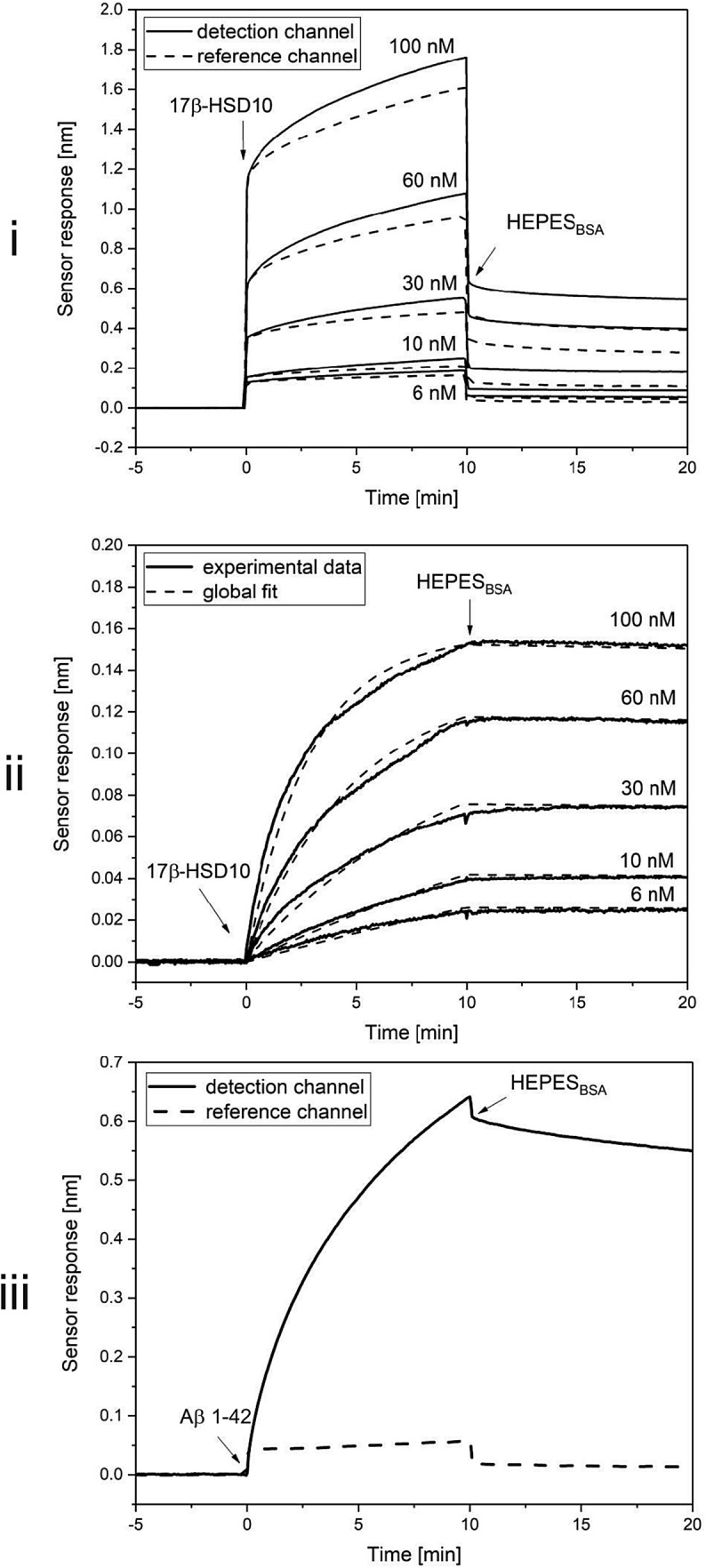
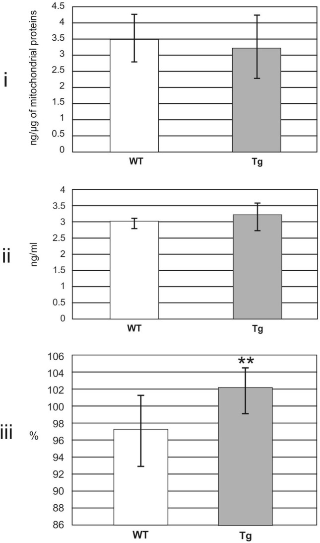
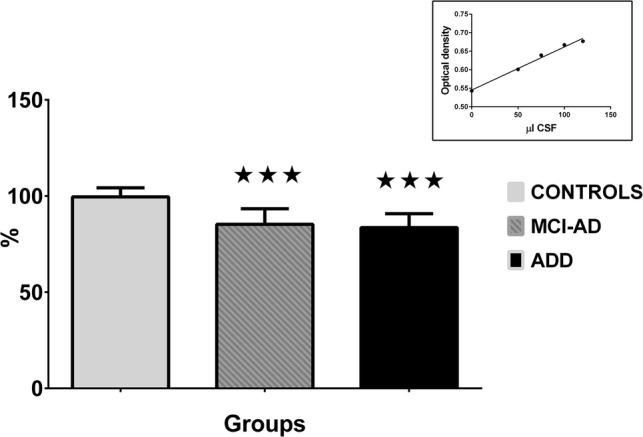
The nucleus-encoded 17β-hydroxysteroid dehydrogenase type 10 (17β-HSD10) regulates cyclophilin D (cypD) in the mitochondrial matrix. CypD regulates opening of mitochondrial permeability transition pores. Both mechanisms may be affected by amyloid β peptides accumulated in mitochondria in Alzheimer's disease (AD). In order to clarify changes occurring in brain mitochondria, we evaluated interactions of both mitochondrial proteins in vitro (by surface plasmon resonance biosensor) and detected levels of various complexes of 17β-HSD10 formed in vivo (by sandwich ELISA) in brain mitochondria isolated from the transgenic animal model of AD (homozygous McGill-R-Thy1-APP rats) and in cerebrospinal fluid samples of AD patients. By surface plasmon resonance biosensor, we observed the interaction of 17β-HSD10 and cypD in a direct real-time manner and determined, for the first time, the kinetic parameters of the interaction (ka 2.0 × 10⁵ M1s-1, kd 5.8 × 10⁴ s-1, and KD 3.5 × 10⁻¹⁰ M). In McGill-R-Thy1-APP rats compared to controls, levels of 17β-HSD10-cypD complexes were decreased and those of total amyloid β increased. Moreover, the levels of 17β-HSD10-cypD complexes were decreased in cerebrospinal fluid of individuals with AD (in mild cognitive impairment as well as dementia stages) or with Frontotemporal lobar degeneration (FTLD) compared to cognitively normal controls (the sensitivity of the complexes to AD dementia was 92.9%, that to FTLD 73.8%, the specificity to AD dementia equaled 91.7% in a comparison with the controls but only 26.2% with FTLD). Our results demonstrate the weakened ability of 17β-HSD10 to regulate cypD in the mitochondrial matrix probably via direct effects of amyloid β. Levels of 17β-HSD10-cypD complexes in cerebrospinal fluid seem to be the very sensitive indicator of mitochondrial dysfunction observed in neurodegeneration but unfortunately not specific to AD pathology. We do not recommend it as the new biomarker of AD.

Keywords: Alzheimer's disease; Amyloid β; Cerebrospinal fluid; Frontotemporal lobar degeneration; Mitochondrial matrix proteins; Transgenic rat model.


Figures

Fig. 1
Fig. 1
Data of SPR biosensor demonstrating the binding of 17β-HSD10 to cypD. Figure demonstrates: (i) the sensor response to binding of 6, 10, 30, 60, and 100 nM 17β-HSD10 in the detection (solid lines) and in the reference (dashed lines) channels, (ii) reference-compensated binding curves for the set of the used concentrations of 17β-HSD10 (solid lines) and their global fits (dashed lines) and finally (iii) the sensor response to the binding of Aβ 1-42 in the detection (solid line) and in the reference (dashed line) channel
Fig. 2
Fig. 2
Demonstration of 17β-HSD10–cypD interaction by co-immunoprecipitation. Figure demonstrates: (i) immunoblot of 17β-HSD10 standard (cca 1 µg), (ii) co-immunoprecipitation of 17β-HSD10–cypD mix pulled-down by anti-cypD antibody-Protein A-sepharose and revealed by anti-17β-HSD10 antibody, (iii) immunoblot of cypD standard (cca 2 µg), and finally (iv) co-immunoprecipitation of 17βHSD10–cypD mix pulled-down by anti-17β-HSD10 antibody-Protein A-sepharose and revealed by anti-cypD antibody
Fig. 3
Fig. 3
Pictures of isolated mitochondria by transmission electron microscopy. Transmission electron microscopy (Imaging Methods Core Facility, BIOCEV, Prague) documented high purity and intactness of mitochondria isolated in accordance with the study of Rajapakse et al. [27]. Sample (mitochondria isolated from three 7-month old male Wistar rats used as internal standards) was stored in 0.25 M sucrose, 0.5 mM K+-EDTA, 10 mM Tris, pH 7.4 with 10% dimethyl sulfoxide at − 80 °C (in concentration of 5 mg protein/ml).
Fig. 4
Fig. 4
Levels of 17β-HSD10, cypD and Aβ in rat mitochondrial fraction estimated by ELISA. Data are presented as the mean ± SD. All values were obtained from 10 WT and 10 Tg (homozygous McGill-R-Thy1-APP) rats. Figure demonstrates: (i) levels of 17β-hydroxysteroid dehydrogenase type 10 (17β-HSD10) estimated by quantitative competitive ELISA (differences between both groups were not statistically significant, Welch test: p = 0.508), (ii) levels of cyclophilin D (cypD) estimated by quantitative sandwich ELISA (differences between both groups were not statistically significant, Welch test: p = 0.091), and finally (iii) total levels of amyloid β (Aβ) peptides estimated by semi-quantitative ELISA (differences between both groups were statistically significant, Welch test: p = 0.006)
Fig. 5
Fig. 5
Levels of 17β-HSD10–cypD and 17β-HSD10–Aβ complexes in rat mitochondrial fraction estimated by semi-quantitative sandwich ELISA. Data are presented as the mean ± SD. Levels of two types of complexes, i.e. of 17β-hydroxysteroid dehydrogenase type 10 and cyclophilin D (17β-HSD10–cypD complexes, experiment (i) and of 17β-hydroxysteroid dehydrogenase type 10 and amyloid β (17β-HSD10–Aβ complexes, experiment (ii) were obtained from 10 WT and 10 Tg (homozygous McGill-R-Thy1-APP) rats. The values of 17β-HSD10–cypD complexes were significantly decreased in Tg compared to WT rats (Welch test: p = 0.012). On the contrary, no differences were found in levels of 17β-HSD10–Aβ complexes (Welch test: p = 0.072)
Fig. 6
Fig. 6
Levels of 17β-HSD10–cypD complexes in CSF of people with AD estimated by semi-quantitative sandwich ELISA. Data (a part of results of Table 2) are presented as the mean ± SD. Levels of complexes were estimated in CSF samples of controls (n = 12), of people with mild cognitive impairment due to Alzheimer's disease (MCI-AD, n = 44) and of people with Alzheimer's disease dementia (ADD, n = 56). Statistical significance (Bonferroni adjusted t-test, separate variance) was calculated with respect to controls (***Adjusted p < 0.001)

Similar articles

Cited by

References

    1. Yang SY, He XY, Schulz H. Multiple functions of type 10 17β-hydroxysteroid dehydrogenase. Trends Endocrin Met. 2005;16:167–175. doi: 10.1016/j.tem.2005.03.006. - DOI - PubMed
    1. Yang SY, He XY, Isaacs C, Dobkin C, Miller D, Philipp M. Roles of 17β-hydroxysteroid dehydrogenase type 10 in neurodegenerative disorders. J Steroid Biochem Mol Biol. 2014;143:460–472. doi: 10.1016/j.jsbmb.2014.07.001. - DOI - PubMed
    1. Rauschenberger K, Schöler K, Sass JO, Sauer S, Djuric Z, Rumig C, Wolf NI, Okun JG, Kolker S, Schwarz H, Fisher C, Grziwa B, Runz H, Numann A, Shafqat N, Kavanagh KL, Hammerling G, Wanders RJA, Shield JPH, Wendel U, Stern D, Nawroth P, Hoffmann GF, Bartram CR, Arnold B, Bierhaus A, Oppermann U, Steinbeisser H, Zschocke J. A non-enzymatic function of 17β-hydroxysteroid dehydrogenase type 10 is required for mitochondrial integrity and cell survival. EMBO Mol Med. 2010;2:51–62. doi: 10.1002/emmm.200900055. - DOI - PMC - PubMed
    1. Harbauer AB, Zahedi RP, Sickman A, Pfanner N, Meisinger C. The protein import machinery of mitochondria—a regulatory hub in metabolism, stress, and disease. Cell Metab. 2014;19:357–372. doi: 10.1016/j.cmet.2014.01.010. - DOI - PubMed
    1. Bertolin G, Jacoupy M, Traver S, Ferrendo-Miguel R, Saint Georges T, Grenier K, Ardila-Osorio H, Muriel MP, Takahashi H, Lees AJ, Gautier C, Guedin D, Coge F, Fon EA, Brice A, Corti O. Parkin maintains mitochondrial levels of the protective Parkinson's disease-related enzyme 17-β hydroxysteroid dehydrogenase type 10. Cell Death Differ. 2015;22:1563–1576. doi: 10.1038/cdd.2014.224. - DOI - PMC - PubMed

Substances

Grants and funding