. 2020 Oct;108(4):545-552.
doi: 10.1007/s10266-020-00486-z. Epub 2020 Jan 30.

Acute cytotoxic effects of silica microparticles used for coating of plastic blood-collection tubes on human periosteal cells

Affiliations

Acute cytotoxic effects of silica microparticles used for coating of plastic blood-collection tubes on human periosteal cells

Hideo Masuki et al. Odontology. 2020 Oct.

Abstract

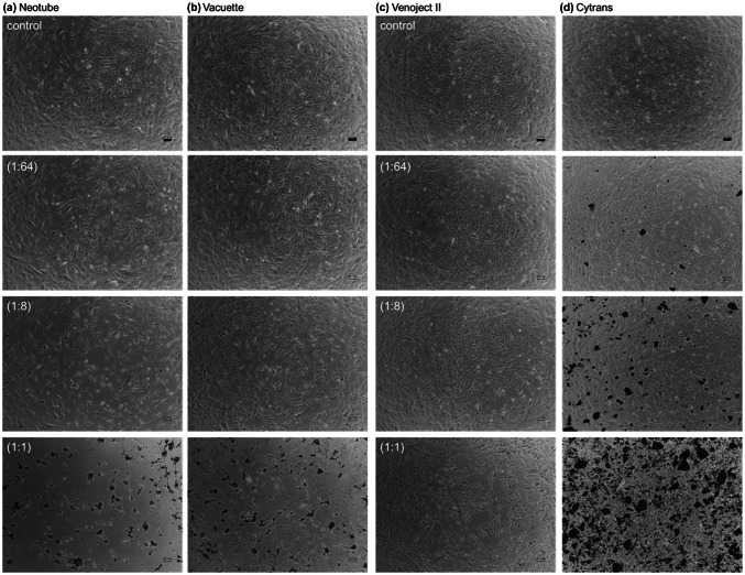
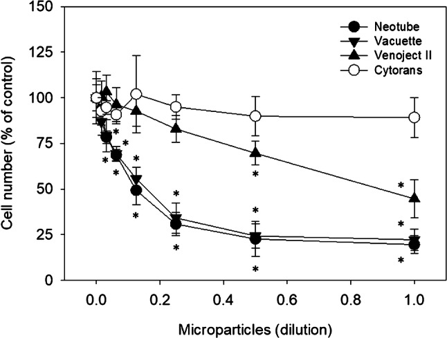
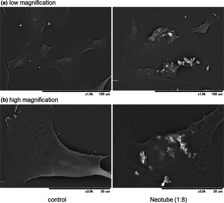
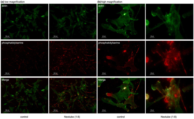
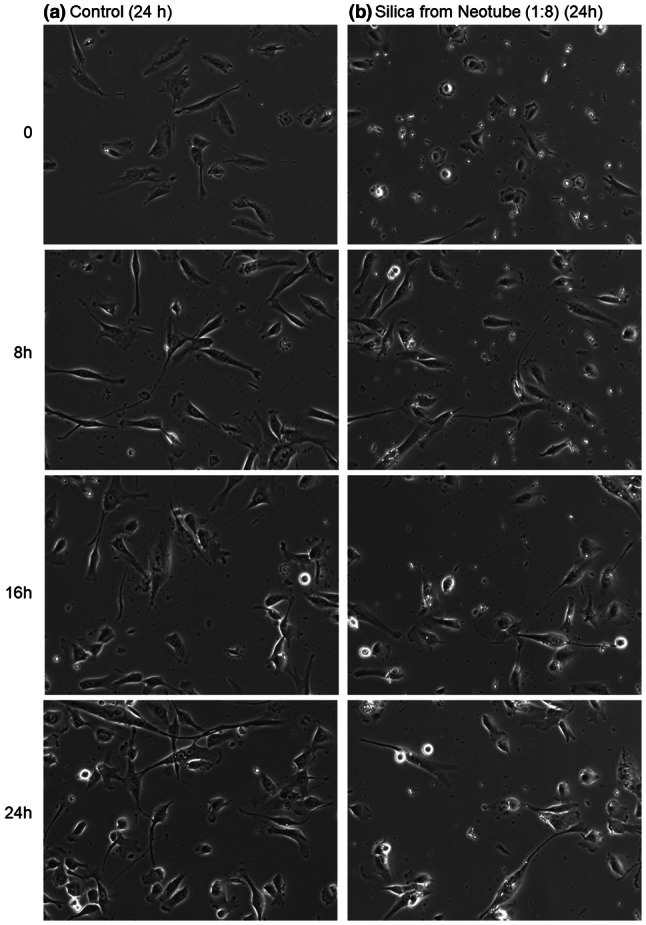
Because of its simple operation, platelet-rich fibrin (PRF) is becoming more popular than the original form, platelet-rich plasma (PRP), in regenerative dentistry. PRF preparation requires plain glass blood-collection tubes, but not either anticoagulants or coagulation factors. However, such glass tubes designed for laboratory testing are no longer commercially available. Although several glass tubes specifically designed for PRF preparation are available, many clinicians prefer to obtain stably supplied substitutes, such as silica-coated plastic tubes produced by major medical device companies. The quality of PRF prepared by silica-coated tubes has not been assessed and we previously reported significant contamination of silica microparticles in the resulting PRF matrix and alerted clinicians against the use for PRF preparation. To further assess the biosafety of the silica microparticles, we presently examined their effects on human normal periosteal cells derived from alveolar bone. The periosteal cells were obtained from explant cultures of small periosteal tissues obtained from healthy donors. Silica microparticles were obtained from silica-coated tubes and added to cell cultures. Cellular responses were monitored using a tetrazolium assay, phase-contract inverted microscopy, an immunofluorescence method, and scanning electron microscopy. Silica microparticles adsorbed onto the cell surface with seemingly high affinity and induced apoptosis, resulting in significant reduction of cell proliferation and viability. These findings suggest that silica microparticles contained in plastic tubes for the purpose of blood coagulation are hazardous for various cell types around sites where silica-contaminated PRF matrices are implanted.

Keywords: Apoptosis; Cytotoxicity; Periosteal cells; Platelet-rich fibrin; Silica.


Conflict of interest statement

The authors declare that they have no conflict of interest.

Figures

Fig. 1
Fig. 1
Phase-contrast images of human periosteal cells treated with silica microparticles. The cells were treated with silica microparticles derived from a Neotube, b Vacuette, or c Venoject II for 72 h. Cells were photographed without fixation. As the negative control, cells were treated with d synthetic carbonate apatite particles (Cytrans Granules). Values in parentheses represent dilution. Scale bar is 100 µm
Fig. 2
Fig. 2
Effects of silica microparticles on the proliferation and viability of human periosteal cells. The cells were seeded into wells of 24-well plates and treated with silica microparticles for 72 h. Cell numbers were assessed using a cell counting kit-8, and the absorbance was measured at 450 nm. Data were obtained from six samples (N = 6) of two representative experiments using periosteal cells derived from two independent donors. Asterisks represent P < 0.05 compared with the negative control, Cytrans Granules
Fig. 3
Fig. 3
Microstructural images of human periosteal cells treated with silica microparticles. The cells were treated with silica microparticles derived from Neotube (1:8 dilution) for 24 h, fixed, and examined using SEM at a low magnification and b high magnification. Similar observations were obtained from four other independent experiments, including Vacuette’s silica, using periosteal cells derived from different donors
Fig. 4
Fig. 4
Fluorescence visualization of apoptosis in human periosteal cells treated with silica microparticles. The cells were treated with silica microparticles derived from Neotube for 24 h. The fixed cells were probed with PE-conjugated Annexin V for detection of phosphatidylserine on cell surface, which is accepted as a marker of apoptosis at a low magnification and b high magnification. The cells were counterstained with FITC-conjugated phalloidin to visualize cytoskeletal polymerized actin. Similar observations were obtained from four other independent experiments, including Vacuette’s silica, using periosteal cells derived from different donors
Fig. 5
Fig. 5
Effects of silica microparticles on the migration and density of human periosteal cells. The cells were treated without silica microparticles derived from Neotube and subjected to time-lapse recording for 24 h. Photomicrographs acquired at 0, 8, 16, and 24 h are shown. Similar observations were obtained from four other independent experiments, including Vacuette’s silica, using periosteal cells derived from different donors

References

    1. Kobayashi M, Kawase T, Horimizu M, et al. A proposed protocol for the standardized preparation of PRF membranes for clinical use. Biologicals. 2012;40:323–329. doi: 10.1016/j.biologicals.2012.07.004. - DOI - PubMed
    1. Tsujino T, Takahashi A, Yamaguchi S, et al. Evidence for contamination of silica microparticles in advanced platelet-rich fibrin matrices prepared using silica-coated plastic tubes. Biomedicines. 2019;7:45. doi: 10.3390/biomedicines7020045. - DOI - PMC - PubMed
    1. Takahashi A, Tsujino T, Yamaguchi S, et al. Distribution of platelets, TGFβ1, PDGF-BB, VEGF, MMP9 and fibronectin in advanced platelet-rich fibrin (A-PRF) and concentrated growth factors (CGF) matrices. J Invest Clin Dent. 2019;10:e12458. doi: 10.1111/jicd.12458. - DOI - PubMed
    1. Margolis J. Glass surface and blood coagulation. Nature. 1956;178:805–806. doi: 10.1038/178805b0. - DOI - PubMed
    1. Finkelstein MM. Silica, silicosis, and lung cancer: a risk assessment. Am J Ind Med. 2000;38:8–18. doi: 10.1002/1097-0274(200007)38:1<8::AID-AJIM2>3.0.CO;2-#. - DOI - PubMed