. 2020 Apr;87(4):633-645.
doi: 10.1002/ana.25691. Epub 2020 Feb 8.

CHIT1 at Diagnosis Reflects Long-Term Multiple Sclerosis Disease Activity

Affiliations

CHIT1 at Diagnosis Reflects Long-Term Multiple Sclerosis Disease Activity

Emanuela Oldoni et al. Ann Neurol. 2020 Apr.

Abstract

Objective: Evidence for a role of microglia in the pathogenesis of multiple sclerosis (MS) is growing. We investigated association of microglial markers at time of diagnostic lumbar puncture (LP) with different aspects of disease activity (relapses, disability, magnetic resonance imaging parameters) up to 6 years later in a cohort of 143 patients.

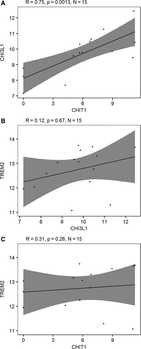
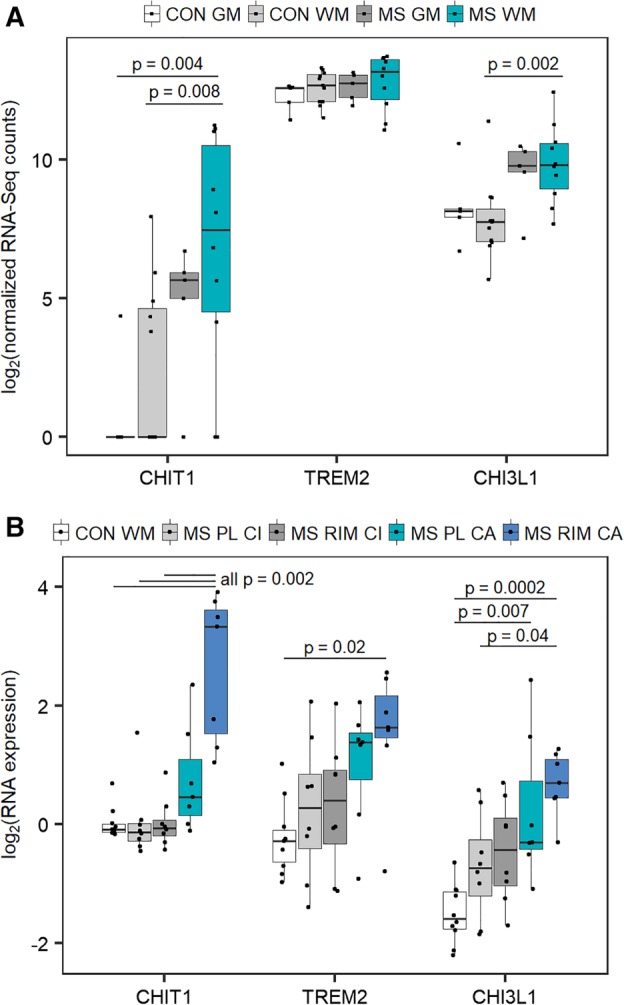
Methods: In cerebrospinal fluid (CSF), we measured 3 macrophage and microglia-related proteins, chitotriosidase (CHIT1), chitinase-3-like protein 1 (CHI3L1 or YKL-40), and soluble triggering receptor expressed on myeloid cells 2 (sTREM2), as well as a marker of neuronal damage, neurofilament light chain (NfL), using enzyme-linked immunosorbent assay and electrochemiluminescence. We investigated the same microglia-related markers in publicly available RNA expression data from postmortem brain tissue.

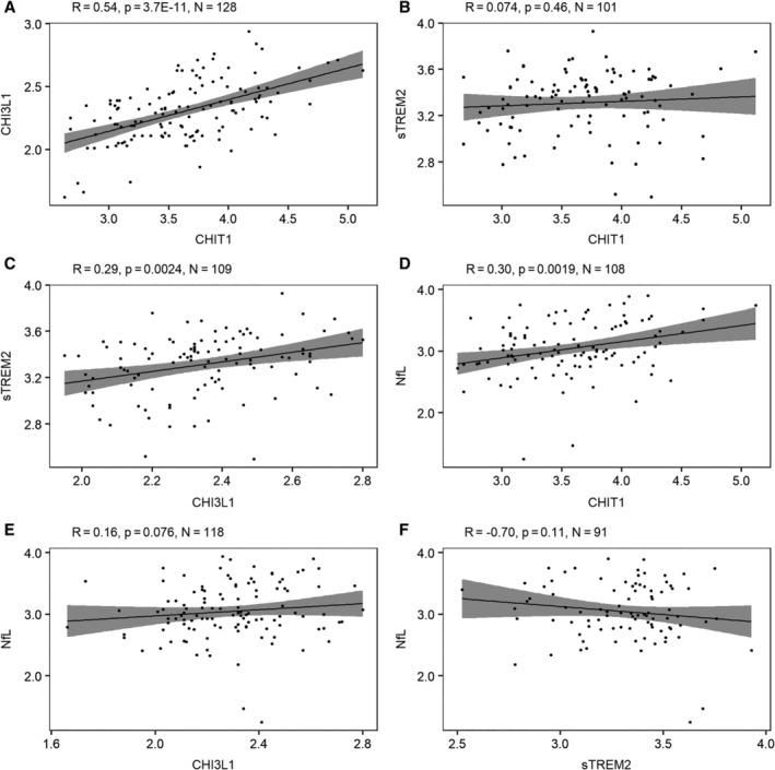
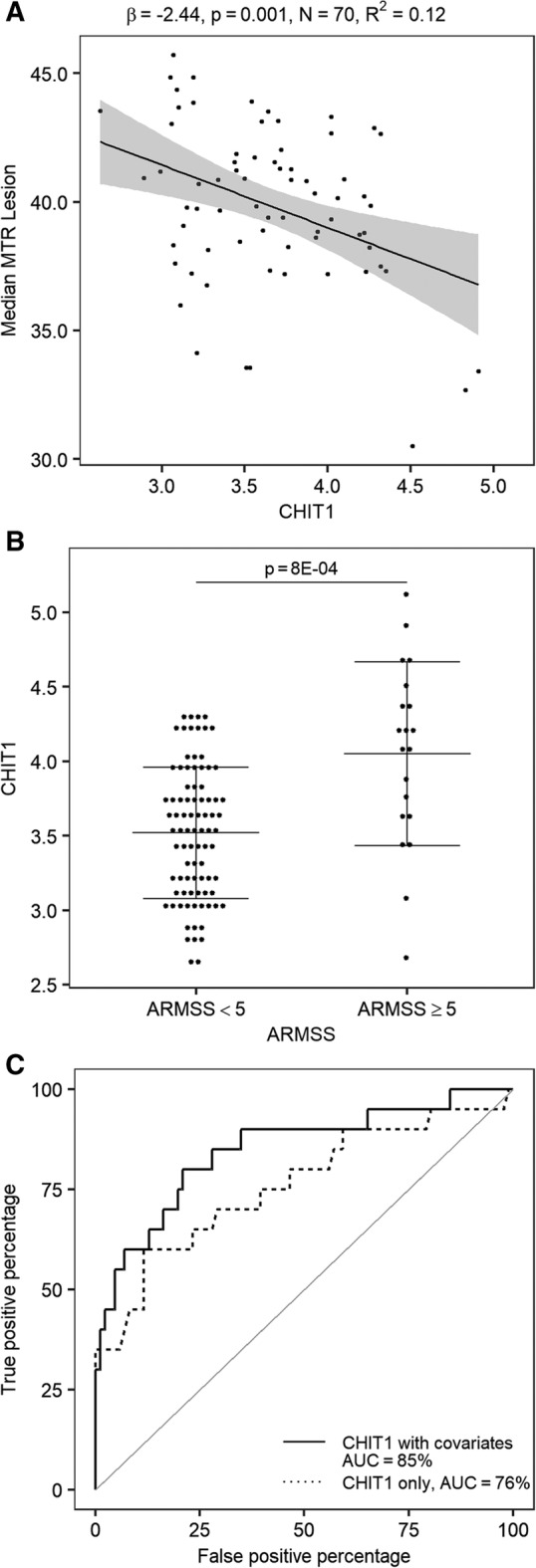
Results: CHIT1 levels at diagnostic LP correlated with 2 aspects of long-term disease activity after correction for multiple testing. First, CHIT1 increased with reduced tissue integrity in lesions at a median 3 years later (p = 9.6E-04). Second, CHIT1 reflected disease severity at a median 5 years later (p = 1.2E-04). Together with known clinical covariates, CHIT1 levels explained 12% and 27% of variance in these 2 measures, respectively, and were able to distinguish slow and fast disability progression (area under the curve = 85%). CHIT1 was the best discriminator of chronic active versus chronic inactive lesions and the only marker correlated with NfL (r = 0.3, p = 0.0019). Associations with disease activity were, however, independent of NfL.

Interpretation: CHIT1 CSF levels measured during the diagnostic LP reflect microglial activation early on in MS and can be considered a valuable prognostic biomarker for future disease activity. ANN NEUROL 2020;87:633-645.


Conflict of interest statement

Nothing to report.

Figures

Figure 1
Figure 1
Correlation between cerebrospinal fluid (CSF) microglial and axonal biomarkers. Pairwise comparisons between CSF markers: (A) chitotriosidase (CHIT1; pg/ml) versus chitinase‐3–like protein 1 (CHI3L1; ng/ml), (B) CHIT1 (pg/ml) versus soluble triggering receptor expressed on myeloid cells 2 (sTREM2; pg/ml), (C) CHI3L1 (ng/ml) versus sTREM2 (pg/ml), (D) CHIT1 (pg/ml) versus neurofilament light chain (NfL; pg/ml), (E) CHI3L1 (ng/ml) versus NfL (pg/ml), and (F) sTREM2 (pg/ml) versus NfL (pg/ml). Probability values are derived from Pearson test. Gray shading shows the 95% confidence bands. The logarithmic (base 10) transformation was applied for all biomarkers.
Figure 2
Figure 2
Correlations between microglial marker RNA expression in brain tissue (corpus callosum) of multiple sclerosis patients.6 Pairwise comparisons of gene expression extracted from publicly available data deposited in the Gene Expression Omnibus database originating from Van der Poel et al6: (A) chitotriosidase (CHIT1) versus chitinase‐3–like protein 1 (CHI3L1), (B) CHI3L1 versus triggering receptor expressed on myeloid cells 2 (TREM2), and (C) CHIT1 versus TREM2. Raw values of RNA‐seq counts were replaced with (value +1) because of the presence of zero values before taking the logarithmic transformation with base 2 as conventional in RNA expression data. Probability values are derived from Pearson test. Gray shading shows the 95% confidence bands.
Figure 3
Figure 3
Association of cerebrospinal fluid chitotriosidase (CHIT1) at diagnosis with primary outcome disease activity parameters at follow‐up. (A) Linear regression analysis for CHIT1 (pg/ml) at diagnostic lumbar puncture (LP) versus median lesion magnetization transfer ratio (MTR; untransformed data, P Shapiro–Wilk = 0.03) at a median 3 years (interquartile range [IQR] = 2–5) later including age at magnetic resonance imaging as covariate; gray shading shows the 95% confidence bands. (B) CHIT1 (pg/ml) at diagnostic LP between groups of fast (Age‐Related Multiple Sclerosis Severity Score [ARMSS] ≥ 5) and slow (ARMSS <5) disability progression as measured at a median 5 years (IQR = 2–7) later. Probability value is derived from a logistic regression including known covariates for disability (sex, age at onset, and disease course); horizontal lines indicate mean and error bars. (C) Receiver operating characteristic curves based on CHIT1 alone or CHIT1 including covariates for discrimination of disability progression as defined in (B). The logarithmic (base 10) transformation was applied for CHIT1.
Figure 4
Figure 4
Correlation between chitotriosidase (CHIT1) 24bp duplication (rs150192398) and CHIT1 levels. X‐axis depicts copies of the minor allele (24bp duplication), which was previously demonstrated to affect RNA stability27 and protein levels.29 The logarithmic transformation (base 10) was applied for CHIT1 (pg/ml) levels. Probability value is derived from linear regression model of allele dosage. 0 = homozygous major allele, n = 87; 1 = heterozygous, n = 34. Horizontal lines indicate mean and error bars.
Figure 5
Figure 5
(A) Microglial marker RNA expression in multiple sclerosis (MS) white and gray matter in 15 MS patients and 10 controls. Probability values are derived from nonparametric Kruskal–Wallis test with Dunn post hoc test on publicly available data deposited in the Gene Expression Omnibus database originating from Van der Poel et al.6 Comparisons without p indicated were not significant. Raw values of RNA‐seq counts were replaced with (value +1) because of the presence of zero values before taking the logarithmic transformation with base 2 as conventional in RNA expression data. (B) Microglial marker RNA expression in active and inactive chronic lesions. Probability values are derived from nonparametric Kruskal–Wallis test with Dunn post hoc test on publicly available data deposited in the Gene Expression Omnibus database originating from Hendrickx et al.5 CHIT1 = chitotriosidase; CHI3L1 = chitinase‐3–like protein 1; CON GM = gray matter control; CON WM = white matter control; MS GM = gray matter from MS patients; MS WM = white matter lesions from MS patients; MS PL CA = perilesion of chronic active lesions from MS patients; MS PL CI = perilesion of chronic inactive lesions from MS patients; MS RIM CA = rim of chronic active lesions from MS patients; MS RIM CI = rim of chronic inactive lesions from MS patients; TREM2 = triggering receptor expressed on myeloid cells 2.

References

    1. Compston A, Coles A. Multiple sclerosis. Lancet 2008;372:1502–1517. - PubMed
    1. Giovannoni G, Bermel R, Phillips T, Rudick R. A brief history of NEDA. Mult Scler Relat Disord 2018;20:228–230. - PubMed
    1. Comabella M, Montalban X. Body fluid biomarkers in multiple sclerosis. Lancet Neurol 2014;13:113–126. - PubMed
    1. Khalil M, Teunissen CE, Otto M, et al. Neurofilaments as biomarkers in neurological disorders. Nat Rev Neurol 2018;14:577–589. - PubMed
    1. Hendrickx DAE, van Scheppingen J, van der Poel M, et al. Gene expression profiling of multiple sclerosis pathology identifies early patterns of demyelination surrounding chronic active lesions. Front Immunol 2017;8:1810. - PMC - PubMed

Publication types