. 2020 Apr;14(4):795-807.
doi: 10.1002/1878-0261.12644. Epub 2020 Feb 20.

RACK1 promotes cancer progression by increasing the M2/M1 macrophage ratio via the NF-κB pathway in oral squamous cell carcinoma

Affiliations

RACK1 promotes cancer progression by increasing the M2/M1 macrophage ratio via the NF-κB pathway in oral squamous cell carcinoma

Hongxia Dan et al. Mol Oncol. 2020 Apr.

Abstract

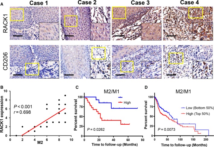
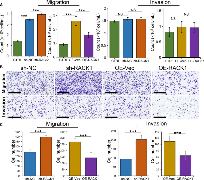
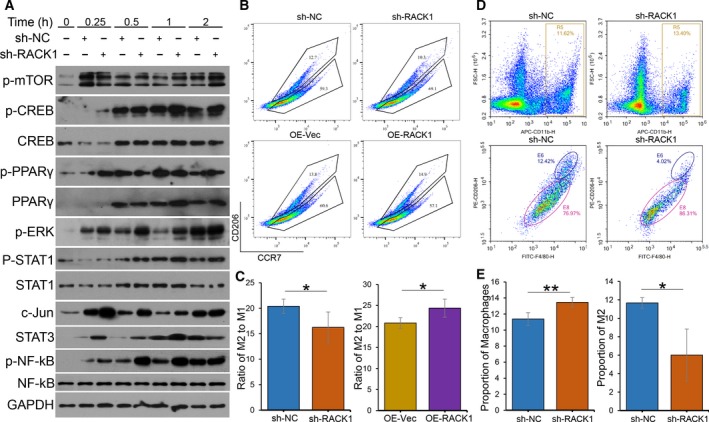
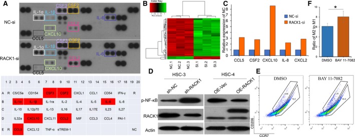
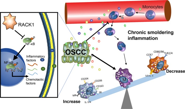
Receptor for activated C kinase 1 (RACK1) has been shown to promote oral squamous cell carcinoma (OSCC) progression, and RACK1 expression levels have been negatively correlated with prognosis in patients with OSCC. Here, we investigated the impact of RACK1 OSCC expression on the recruitment and differentiation of tumor-associated macrophages. High RACK1 expression in OSCC cells correlated with increased M2 macrophage infiltration in tumor samples from a clinical cohort study. Moreover, the combination of RACK1 expression and the M2/M1 ratio could successfully predict prognosis in OSCC. OSCC cells with high RACK1 expression inhibited the migration of THP-1 cells, promoted M2-like macrophage polarization in vitro, and increased the proportion of M2-like macrophages in a xenograft mouse model. Moreover, both M1- and M2-like macrophage polarization-associated proteins were induced in macrophages cocultured with RACK1-silenced cell supernatant. A mechanistic study revealed that the expression and secretion of C-C motif chemokine 2 (CCL2), C-C motif chemokine 5 (CCL5), interleukin-6 (IL-6), and interleukin-1 (IL-1) are closely related to RACK1 expression. In addition, blocking nuclear factor-kappa B (NF-κB) could promote M2-like macrophage polarization. These results indicate that RACK1 and the M2/M1 ratio are predictors of a poor prognosis in OSCC. RACK1 promotes M2-like polarization by regulating NF-κB and could be used as a potential therapeutic target for antitumor immunity.

Keywords: NF-κB; RACK1; macrophage polarization; oral squamous cell carcinoma; tumor-associated macrophages.


Conflict of interest statement

The authors declare no conflicts of interest.

Figures

Figure 1
Figure 1
The M2/M1 ratio is positively correlated with the RACK level and is associated with a poor prognosis in OSCC. (A) IHC staining of 36 paraffin‐embedded OSCC sections with human antibodies against RACK1 and CD206 (scale bar: 100 μm). (B) Statistical analysis revealed that the expression intensity of RACK was positively correlated with the number of M2 macrophages (r = 0.698, P < 0.001). (C) Kaplan–Meier survival analysis revealed that a high M2/M1 ratio indicated a poor OS in 36 OSCC patients (P = 0.0262). (D) TCGA database analysis showed that a high M2/M1 ratio indicated a poor OS in human primary oral cancers (P < 0.01).
Figure 2
Figure 2
RACK1 inhibits the capacity of OSCC cells to recruit macrophages. (A) Transwell migration and Transwell Matrigel invasion assays used THP‐1 cells in the upper chamber and infected OSCC supernatants in the lower chamber. The average numbers of migrated and invaded cells were quantified (mean ± SD; ***P < 0.001). (B) Transwell migration and Transwell Matrigel invasion assays used RAW264.7 cells in the upper chamber and infected OSCC supernatants in the lower chamber (scale bars: 200 μm). (C) The average numbers of migrated and invaded RAW264.7 cells were quantified (mean ± SD; ***P < 0.001).
Figure 3
Figure 3
RACK1 inhibits macrophage activation but increases the proportion of M2 macrophages. (A) Immunoblots of p‐mTOR, CREB, p‐CREB, PPARγ, p‐PPARγ, p‐ERK, STAT1, p‐STAT1, c‐Jun, STAT3, NF‐κB and p‐NF‐κB in macrophages, induced from THP‐1 cells by 100 ng·mL−1 PMA for 24 h, cocultured with infected OSCC supernatants for the indicated times (0, 0.25, 0.5, 1 and 2 h). (B) Macrophages were induced from THP‐1 cells by PMA following incubation with transfected OSCC supernatants (sh‐NC, sh‐RACK1, OE‐Vec or OE‐RACK1) for 8 h. After centrifugation, the macrophages were stained with CD206 and CCR7 antibodies. The percentages and cell numbers of different macrophages were analyzed using flow cytometry. (C) Analysis of the M2/M1 ratio of different THP‐1‐induced groups detected by flow cytometry (mean ± SD; *P < 0.05). (D) Single cells were isolated from tumor tissues using collagenase IV and then stained with CD206, CD11b and F4/80 antibodies. The percentages and cell numbers of macrophages were analyzed using flow cytometry. R5: CD11b‐positive macrophages; E6: both CD206‐ and F4/80‐positive macrophages (M2). (E) Analysis of the numbers of M0 and M2 macrophages in tumor tissues from different groups detected by flow cytometry (mean ± SD; *P < 0.05, **P < 0.01).
Figure 4
Figure 4
RACK1 increases the M2/M1 ratio in an NF‐κB axis‐dependent manner. (A) Human cytokine array analysis of the conditioned medium from HSC‐3 control cells and HSC‐3 cells with RACK1 silencing for 36 h. (B) Unsupervised hierarchical clustering analysis by complete linkage between si‐NC‐ and si‐RACK1‐transfected HSC‐3 cells for 36 h. (C) Analysis of CCL5, CSF2, CXCL10, IL‐8 and CXCL2 mRNA levels in HSC‐3 cells transfected with si‐NC‐ or si‐RACK1 for 36 h (P < 0.05). (D) Western blots for p‐NF‐κB and RACK1 in two infected OSCC cell lines. (E) Macrophages were induced from THP‐1 cells by PMA and cocultured with sh‐NC HSC‐3 cell supernatant (with DMSO or BAY 11‐7082 included) for 8 h. After centrifugation, the macrophages were stained with CD206 and CCR7 antibodies. The percentages and cell numbers of macrophages were analyzed using flow cytometry. (F) Analysis of the M2/M1 ratio in different induction groups (DMSO or BAY 11‐7082) (mean ± SD; *P < 0.05).
Figure 5
Figure 5
Schematic model by which RACK1 promotes the progression of OSCC. RACK1 inhibits the activation of NF‐κB, regulates the expression and secretion of proinflammatory factors and macrophage chemokines, inhibits the massive recruitment of macrophages and severe inflammatory reactions, induces a chronic smoldering inflammation microenvironment and promotes the development of tumors.

Similar articles

Cited by

References

    1. Alves AM, Diel LF and Lamers ML (2018) Macrophages and prognosis of oral squamous cell carcinoma: a systematic review. J Oral Pathol Med 47, 460–467. - PubMed
    1. Balermpas P, Rodel F, Liberz R, Oppermann J, Wagenblast J, Ghanaati S, Harter PN, Mittelbronn M, Weiss C, Rodel C et al (2014) Head and neck cancer relapse after chemoradiotherapy correlates with CD163+ macrophages in primary tumour and CD11b+ myeloid cells in recurrences. Br J Cancer 111, 1509–1518. - PMC - PubMed
    1. Balkwill F, Charles KA and Mantovani A (2005) Smoldering and polarized inflammation in the initiation and promotion of malignant disease. Cancer Cell 7, 211–217. - PubMed
    1. Berns H, Humar R, Hengerer B, Kiefer FN and Battegay EJ (2000) RACK1 is up‐regulated in angiogenesis and human carcinomas. FASEB J 14, 2549–2558. - PubMed
    1. Brown JM, Recht L and Strober S (2017) The promise of targeting macrophages in cancer therapy. Clin Cancer Res 23, 3241–3250. - PMC - PubMed

Publication types

MeSH terms