. 2020 Jan-Dec;12(1):1715705.
doi: 10.1080/19420862.2020.1715705.

Improved translation of stability for conjugated antibodies using an in vitro whole blood assay

Affiliations

Improved translation of stability for conjugated antibodies using an in vitro whole blood assay

Aimee Fourie-O'Donohue et al. MAbs. 2020 Jan-Dec.

Abstract

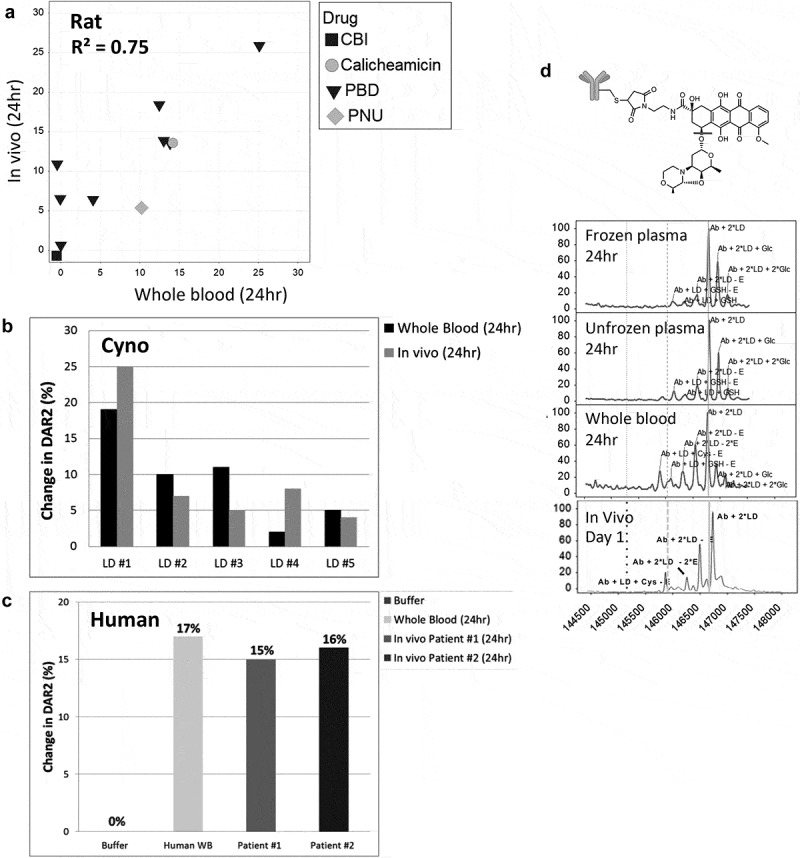
For antibody-drug conjugates to be efficacious and safe, they must be stable in circulation to carry the payload to the site of the targeted cell. Several components of a drug-conjugated antibody are known to influence stability: 1) the site of drug attachment on the antibody, 2) the linker used to attach the payload to the antibody, and 3) the payload itself. In order to support the design and optimization of a high volume of drug conjugates and avoid unstable conjugates prior to testing in animal models, we wanted to proactively identify these potential liabilities. Therefore, we sought to establish an in vitro screening method that best correlated with in vivo stability. While traditionally plasma has been used to assess in vitro stability, our evaluation using a variety of THIOMABTM antibody-drug conjugates revealed several disconnects between the stability assessed in vitro and the in vivo outcomes when using plasma. When drug conjugates were incubated in vitro for 24 h in mouse whole blood rather than plasma and then analyzed by affinity capture LC-MS, we found an improved correlation to in vivo stability with whole blood (R2 = 0.87, coefficient of determination) compared to unfrozen or frozen mouse plasma (R2 = 0.34, 0.01, respectively). We further showed that this whole blood assay was also able to predict in vivo stability of other preclinical species such as rat and cynomolgus monkey, as well as in human. The screening method utilized short (24 h) incubation times, as well as a custom analysis software, allowing increased throughput and in-depth biotransformation characterization. While some instabilities that were more challenging to identify remain, the method greatly enhanced the process of screening, optimizing, and lead candidate selection, resulting in the substantial reduction of animal studies.

Keywords: Stability; antibody-drug conjugate; drug modification; plasma; whole blood.


Figures

Figure 1.
Figure 1.
Lack of in vitro plasma stability translation to in vivo results. Correlation of mouse in vivo stability with in vitro stability for 13 TDC conjugates in frozen mouse plasma (a and b) and unfrozen mouse plasma (c and d) at 24 hr or 96 hr where 0 = no loss (stable) and 100 = complete loss (unstable); “loss” = linker-drug deconjugation and/or inactivating payload metabolism. Axes represent percent loss of drug relative to 0 h. (e, f) Specific examples of mouse plasma stability of MS spectra DAR profiles (top, middle) showing disconnect with in vivo stability (bottom) for an anti-HER2 TDC conjugated with an anthracycline analog (PNU) (e) and an anti-HER2 TDC conjugated to MMAE (f). MS peak labels indicate an antibody (Ab, glycated (Glc)) with one linker drug (+ LD) or two (+ 2*LD) can have drug lost and replaced by a cysteine (+ Cys) or a glutathione (+ GSH) or modified (-E).
Figure 2.
Figure 2.
Improved correlation of in vitro stability with in vivo stability using whole blood. Correlation of TDC in vivo stability with unfrozen whole blood compared to in vivo at 24 h (a) using the same 13 conjugates evaluated in plasma (Figure 1). (b, c) Specific examples of MS spectra of in vivo DAR stability profiles (B, C bottom) compared to stability profile in whole blood (top) for an anti-HER2 TDC conjugated with an anthracycline analog (PNU) (b) and an anti-HER2 TDC conjugated to MMAE (c). Axes in (A) represent the loss of drug percentage relative to 0 hr. MS peak labels indicate an antibody (Ab, glycated (Glc)) with one linker drug (+ LD) or two (+ 2*LD) can have drug lost and replaced by a cysteine (+ Cys) or a glutathione (+ GSH) or modified (-E). A 96 h time-point in whole blood was not evaluated because, after 48 h at 37°C, the samples could not be processed due to clotting of the whole blood.
Figure 3.
Figure 3.
Identification of stability outliers between mouse in vitro whole blood and in vivo outcomes. Additional TDC conjugates were evaluated for stability and their in vitro and in vivo correlation compared (a). TDC conjugates outside of the ± 20% (dashed line) of linear regression line (solid line) were identified as outliers and spectra are shown (B, C, D). Axis in (A) represents a change in DAR2 at 24 h in matrix relative to buffer at 0 hr. MS peak labels indicate an antibody (Ab, glycated (Glc)) with one linker drug (+ LD) or two (+ 2*LD) can have drug lost and replaced by a cysteine (+ Cys) or a glutathione (+ GSH) or modified (acetyl loss – Ac, methyl loss – Me, amide and ester hydrolysis – B).
Figure 4.
Figure 4.
In vitro to in vivo stability translation in other species. Correlation of in vitro TDC stability with in vivo stability in (a) rat (top), (b) cyno, and (c) human (bottom). Evaluated 11 conjugates for rat, 5 pyrrolobenzodiazepine conjugates with different linkers for cyno and 1 conjugate for human. (b) Specific example of a cyno plasma stability disconnect with cyno whole blood and in vivo for a TDC with an anthracycline analog. MS peak labels indicate an antibody (Ab, glycated (Glc)) with one linker drug (+ LD) or two (+ 2*LD) can have drug lost and replaced by a cysteine (+ Cys) or a glutathione (+ GSH) or modified (ether cleavage – E).
Figure 5.
Figure 5.
Reproducibility and species difference of whole blood stability for two control TDCs (a, b) using whole blood shipped overnight (n = 13). Differences in drug modification (b ether cleavage (-E), e carbamate hydrolysis (-CB)) and DAR (c disulfide cleavage, f maleimide cleavage) between buffer and four species for two different conjugates (b, c and e, f) run as controls for each shipment of whole blood.

References

    1. Chari RVJ, Miller ML, Widdison WC.. Antibody–drug conjugates: an emerging concept in cancer therapy. Angew Chem Int Ed Engl. 2014. April 7;53(15):3796–11. doi:10.1002/anie.201307628. - DOI - PubMed
    1. Peters C, Brown S. Antibody–drug conjugates as novel anti-cancer chemotherapeutics. Biosci Rep. 2015. June 12;35(4). doi:10.1042/BSR20150089. - DOI - PMC - PubMed
    1. Wang H, Rangan VS, Sung MC, Passmore D, Kempe T, Wang X, Thevanayagam L, Pan C, Rao C, Srinivasan M, et al. Pharmacokinetic characterization of BMS-936561, an anti-CD70 antibody-drug conjugate, in preclinical animal species and prediction of its pharmacokinetics in humans. Biopharm Drug Dispos. 2016. March;37(2):93–106. doi:10.1002/bdd.1953. - DOI - PubMed
    1. Polakis P. Antibody drug conjugates for cancer therapy. Pharmacol Rev. 2016. January;68(1):3–19. doi:10.1124/pr.114.009373. - DOI - PubMed
    1. Strop P, Delaria K, Foletti D, Witt JM, Hasa-Moreno A, Poulsen K, Casas MG, Dorywalska M, Farias S, Pios A, et al. Site-specific conjugation improves therapeutic index of antibody drug conjugates with high drug loading. Nat Biotechnol. 2015. July;33(7):694–96. doi:10.1038/nbt.3274. - DOI - PubMed

Substances

LinkOut - more resources