. 2020 Jan 4:2020:4717258.
doi: 10.1155/2020/4717258. eCollection 2020.

Ezetimibe Attenuates Oxidative Stress and Neuroinflammation via the AMPK/Nrf2/TXNIP Pathway after MCAO in Rats

Affiliations

Ezetimibe Attenuates Oxidative Stress and Neuroinflammation via the AMPK/Nrf2/TXNIP Pathway after MCAO in Rats

Jing Yu et al. Oxid Med Cell Longev. .

Abstract

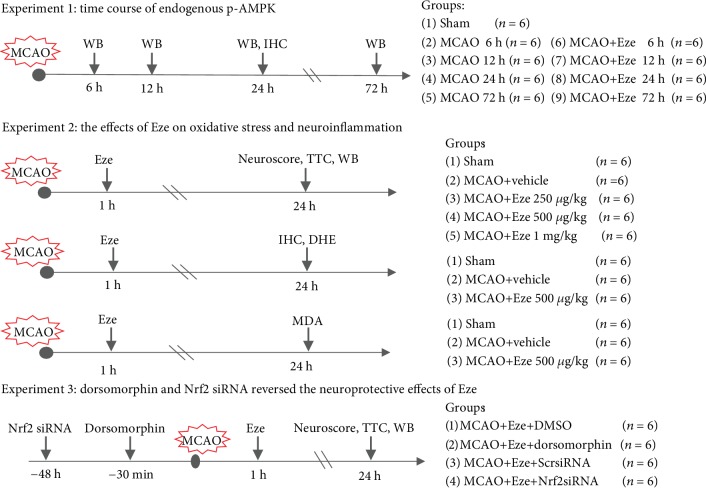
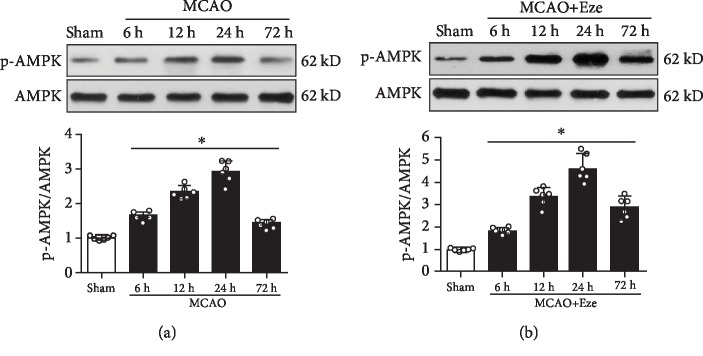
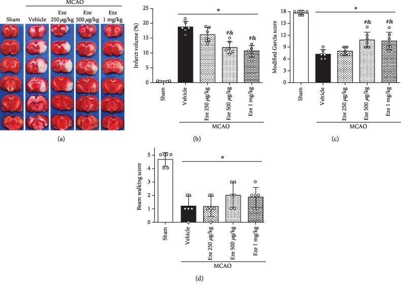
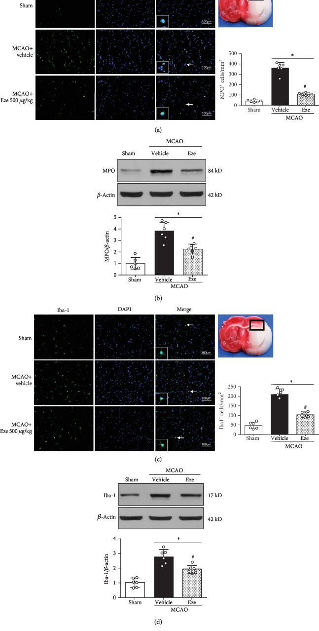
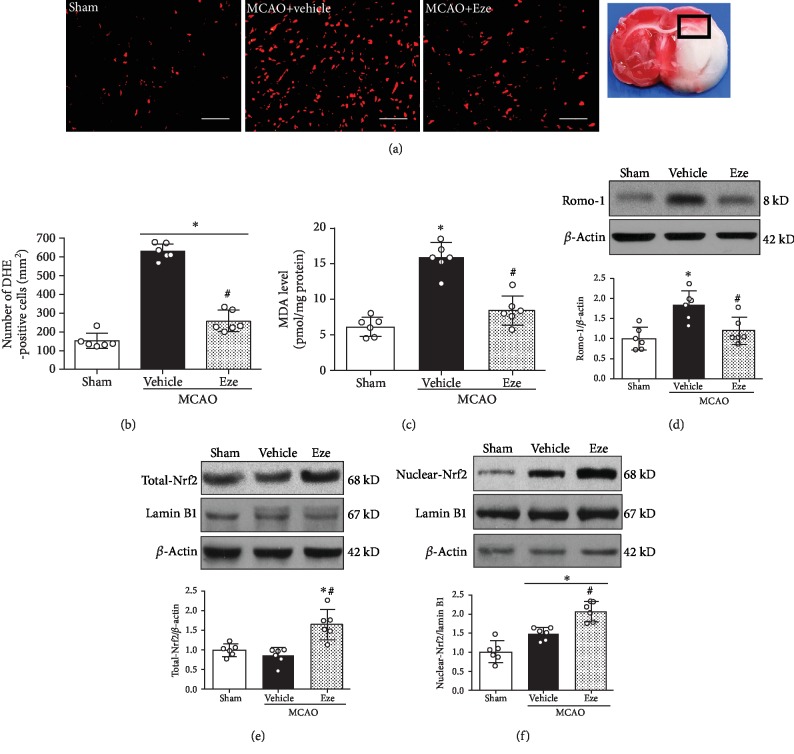
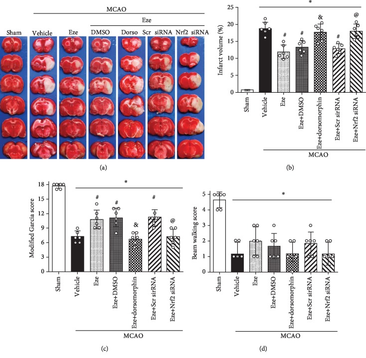
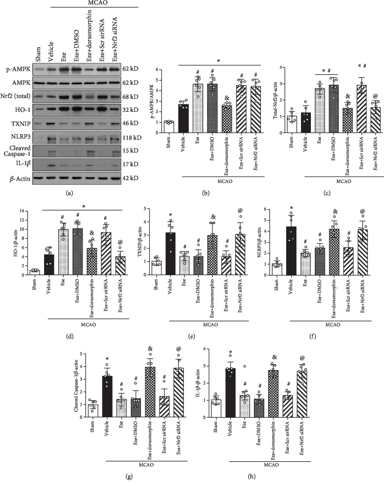
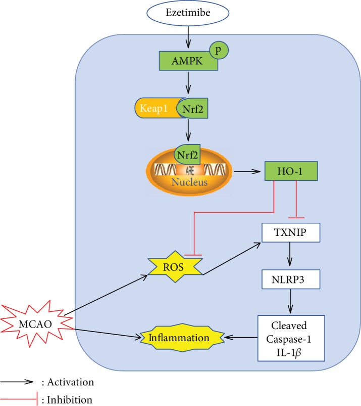
Oxidative stress and neuroinflammation play essential roles in ischemic stroke-induced brain injury. Previous studies have reported that Ezetimibe (Eze) exerts antioxidative stress and anti-inflammatory properties in hepatocytes. In the present study, we investigated the effects of Eze on oxidative stress and neuroinflammation in a rat middle cerebral artery occlusion (MCAO) model. One hundred and ninety-eight male Sprague-Dawley rats were used. Animals assigned to MCAO were given either Eze or its control. To explore the downstream signaling of Eze, the following interventions were given: AMPK inhibitor dorsomorphin and nuclear factor erythroid 2-related factor 2 (Nrf2) siRNA. Intranasal administration of Eze, 1 h post-MCAO, further increased the endogenous p-AMPK expression, reducing brain infarction, neurologic deficits, neutrophil infiltration, microglia/macrophage activation, number of dihydroethidium- (DHE-) positive cells, and malonaldehyde (MDA) levels. Specifically, treatment with Eze increased the expression of p-AMPK, Nrf2, and HO-1; Romo-1, thioredoxin-interacting protein (TXNIP), NOD-like receptor protein 3 (NLRP3), Cleaved Caspase-1, and IL-1β were reduced. Dorsomorphin and Nrf2 siRNA reversed the protective effects of Eze. In summary, Eze decreases oxidative stress and subsequent neuroinflammation via activation of the AMPK/Nrf2/TXNIP pathway after MCAO in rats. Therefore, Eze may be a potential therapeutic approach for ischemic stroke patients.


Conflict of interest statement

The authors declare that they have no conflict of interest.

Figures

Figure 1
Figure 1
Experimental design and animal groups. DHE: dihydroethidium; DMSO: dimethyl sulfoxide; dorsomorphin: AMPK inhibitor; Eze: Ezetimibe; IHC: immunohistochemistry; MCAO: middle cerebral artery occlusion; MDA: malonaldehyde; Scr siRNA: scramble siRNA; TTC: 2,3,5-triphenyltetrazolium chloride; WB: western blot.
Figure 2
Figure 2
Expression of endogenous p-AMPK after MCAO. (a) Representative western blot bands and quantitative analysis of p-AMPK expression in the ipsilateral hemisphere after MCAO. (b) Representative western blot band and quantitative analysis of p-AMPK expression after treatment with Ezetimibe. The dose of Ezetimibe was 500 μg/kg. p < 0.05 vs. sham. n = 6 per group.
Figure 3
Figure 3
Eze reduced brain infraction and improved neurological outcomes at 24 h after MCAO. (a–c) Intranasal administration of Eze reduced infarction volume and improved the Garcia score at 24 h after MCAO. (d) There were no differences in beam walking scores between the MCAO groups. p < 0.05 vs. sham, #p < 0.05 vs. vehicle, and &p < 0.05 vs. Eze 250 μg/kg. n = 6 per group. Eze: Ezetimibe.
Figure 4
Figure 4
Eze inhibited microglia/macrophage activation and neutrophil infiltration at 24 h after MCAO. (a, c) Immunofluorescence revealed that treatment with Eze reduced the number of MPO-positive cells and Iba-1-positive cells in the ischemic penumbra region. (b, d) Representative western blot bands and quantitative analyses of MPO and Iba-1 protein levels at 24 h after MCAO. p < 0.05 vs. sham; #p < 0.05 vs. vehicle. n = 6 per group. Eze: Ezetimibe. Scale bar = 100 μm.
Figure 5
Figure 5
Eze reduced oxidative stress injury at 24 h after MCAO. (a) Representative microphotograph of DHE staining in the ischemic penumbra region. (b) Quantitative analysis of DHE-positive cells in the ischemic penumbra region. (c) The MDA level in the ischemic penumbra region. (d) Representative western blot band and quantitative analysis of Romo-1 expression. (e, f) Representative western blot bands and quantitative analyses of total-Nrf2 and nuclear-Nrf2 expression. p < 0.05 vs. sham; #p < 0.05 vs. vehicle. n = 6 per group. DHE: dihydroethidium; Eze: Ezetimibe; MDA: malonaldehyde. Scale bar = 100 μm.
Figure 6
Figure 6
Dorsomorphin and Nrf2 siRNA reversed the neuroprotective effects of Eze after MCAO. (a) Representative image of TTC-staining brain slices, (b) quantified infarction volumes, (c) modified Garcia scores, and (d) beam walking scores at 24 h after MCAO. p < 0.05 vs. sham, #p < 0.05 vs. vehicle, &p < 0.05 vs. Eze+DMSO, and @p < 0.05 vs. Eze+Scr siRNA. n = 6 per group. Dorso: dorsomorphin; Eze: Ezetimibe; Scr siRNA: scramble siRNA.
Figure 7
Figure 7
Eze attenuated oxidative stress and neuroinflammation via the AMPK/Nrf2/TXNIP pathway after MCAO. (a) Representative western blot bands. (b–h) Quantitative analyses of p-AMPK, Nrf2, HO-1, TXNIP, NLRP3, Cleaved Caspase-1, and IL-1β in the ipsilateral hemisphere at 24 h after MCAO. p < 0.05 vs. sham, #p < 0.05 vs. vehicle, &p < 0.05 vs. Eze+DMSO, and @p < 0.05 vs. Eze+Scr siRNA. n = 6 per group. Eze: Ezetimibe; Scr siRNA: scramble siRNA.
Figure 8
Figure 8
Proposed pathway for Eze and its downstream targets. Eze increases levels of phosphorylated AMPK and activates Nrf2, which translocates into the nucleus to increase the transcription of HO-1—a protein that inhibits ROS and inflammation.

Similar articles

Cited by

References

    1. GBD 2016 Causes of Death Collaborators. Global, regional, and national age-sex specific mortality for 264 causes of death, 1980-2016: a systematic analysis for the Global Burden of Disease Study 2016. The Lancet. 2017;390(10100):1151–1210. doi: 10.1016/S0140-6736(17)32152-9. - DOI - PMC - PubMed
    1. Moretti R., Pansiot J., Bettati D., et al. Blood-brain barrier dysfunction in disorders of the developing brain. Frontiers in Neuroscience. 2015;9:p. 40. doi: 10.3389/fnins.2015.00040. - DOI - PMC - PubMed
    1. Ozkul A., Akyol A., Yenisey C., Arpaci E., Kiylioglu N., Tataroglu C. Oxidative stress in acute ischemic stroke. Journal of Clinical Neuroscience. 2007;14(11):1062–1066. doi: 10.1016/j.jocn.2006.11.008. - DOI - PubMed
    1. Chamorro A., Dirnagl U., Urra X., Planas A. M. Neuroprotection in acute stroke: targeting excitotoxicity, oxidative and nitrosative stress, and inflammation. Lancet Neurology. 2016;15(8):869–881. doi: 10.1016/S1474-4422(16)00114-9. - DOI - PubMed
    1. Shen M. H., Zhang C. B., Zhang J. H., Li P. F. Electroacupuncture attenuates cerebral ischemia and reperfusion injury in middle cerebral artery occlusion of rat via modulation of apoptosis, inflammation, oxidative stress, and excitotoxicity. Evidence-based Complementary and Alternative Medicine. 2016;2016:15. doi: 10.1155/2016/9438650.9438650 - DOI - PMC - PubMed

MeSH terms