. 2019 Oct;22(10):1158-1165.
doi: 10.22038/ijbms.2019.36189.8622.

Filtered Kombucha tea ameliorates the leaky gut syndrome in young and old mice model of colitis

Affiliations

Filtered Kombucha tea ameliorates the leaky gut syndrome in young and old mice model of colitis

Nafiseh Pakravan et al. Iran J Basic Med Sci. 2019 Oct.

Abstract

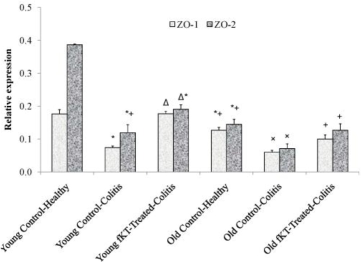
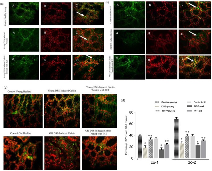
Objectives: Zonula occludens proteins (ZO-1 and ZO-2) are important intracellular tight junction (TJ)-associated proteins that link the cell cytoskeleton to the trans-membrane TJ proteins. Destruction of TJ proteins is called the "leaky gut syndrome" and has been observed in some of the gastrointestinal diseases such as the inflammatory bowel disease (IBD). So, therapeutic approaches aim to restore the expression of TJ proteins and reduce intestinal permeability. Healing effect of Kombucha tea (KT), so-called long-life mushroom, on the gastrointestinal system, particularly its extraordinary healing effects on intestinal ulcers has been purported traditionally and rarely reported scientifically. This study aimed to investigate the therapeutic effect of filtered KT (fKT) in young and old mice model of colitis.

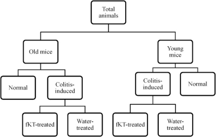
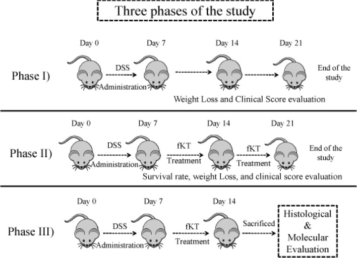
Materials and methods: Leaky gut was induced in two groups of young and old age using dextran sodium sulfate in drinking water for seven days. Then, fKT was administered to the mice affected by colitis and compared with the age-matched normal and untreated animals with colitis.

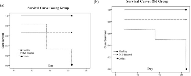
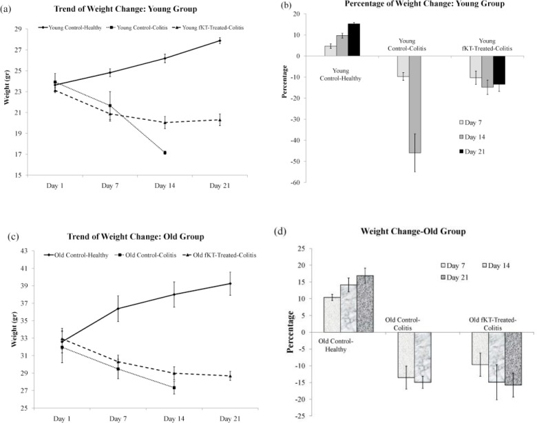
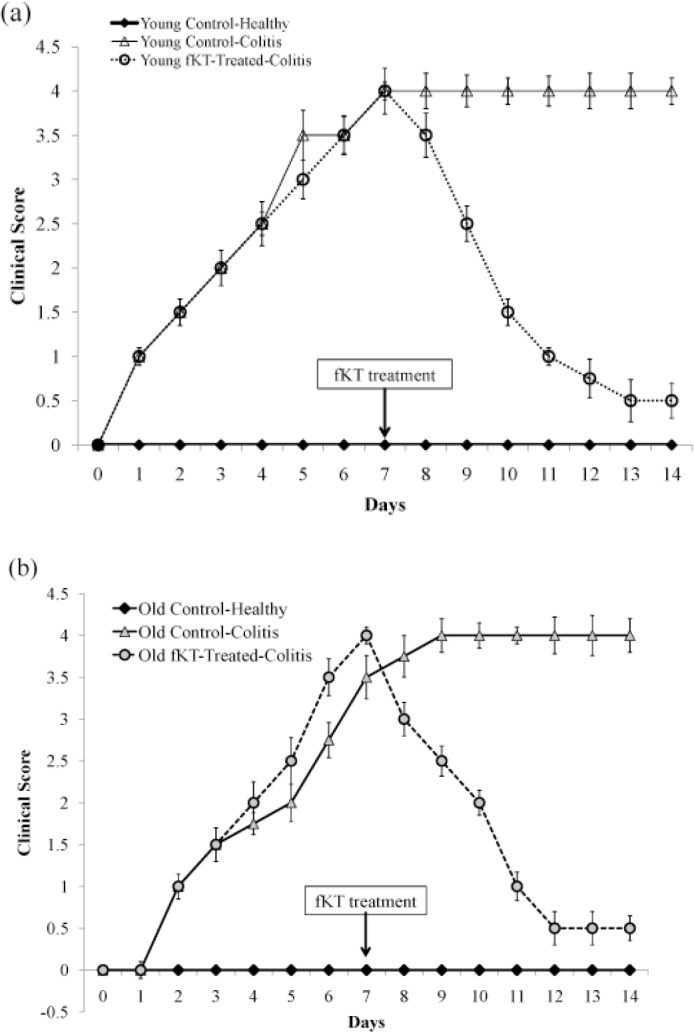
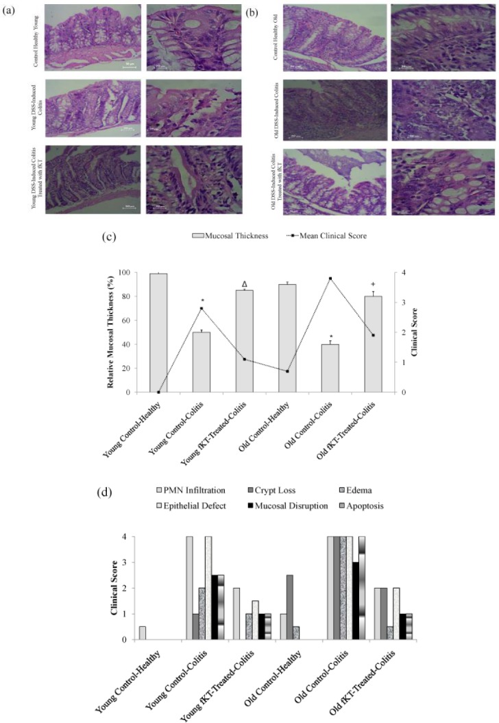
Results: Survival rate of the fKT-treated young and old animals with colitis increased and weight loss decreased. Accordingly, digestive disorders characterized by bleeding and diarrhea were improved in fKT-treated mice. Molecular and histological examination indicated that expression of ZO-1 and ZO-2 was significantly improved in fKT-treated mice.

Conclusion: Our results suggest KT as a promising therapeutic candidate to reduce intestinal permeability. Young animals with colitis showed more severe clinical signs and less survival rate than old mice with colitis, but this group responded better to fKT treatment than the old mice.

Keywords: Age; Colitis; Kombucha tea; Leaky gut; ZO-1; ZO-2.


Conflict of interest statement

The authors have no competing interests.

Figures

Figure 1
Figure 1
Experimental groups of the study
Figure 2
Figure 2
Three phases of the study through which experimental groups were designed and treated
Figure 3
Figure 3
Survival curve of young (a) and old (b) groups including young and old healthy control, young and old DSS-induced colitis, and young and old DSS-induced colitis treated with fKT. Animals were monitored every day for the duration of the experiment. Survival rate was markedly increased by fKT treatment. Analysis made by Kaplan–Meier and compared using the log-rank test
Figure 4
Figure 4
Trend of weight change and percentage of weight loss in the young (a, b) and old (c, d) groups including young and old healthy control, young and old DSS-induced colitis, and young and old DSS-induced colitis treated with fKT. Weight change and weight loss are remarkably affected by fKT treatment
Figure 5
Figure 5
Clinical Score of the young (a) and old (b) DSS-induced colitis mice treated with fKT comparing with the age-matched healthy or DSS-induced colitis mice measured daily from the day of disease induction. In line with the survival rate and weight loss, the clinical score was also improved by fKT treatment. Data are presented as mean±SD
Figure 6
Figure 6
(a) H&E stained colon sections of healthy young animals, young DSS-induced colitis, and young DSS-induced colitis treated with fKT, (b) healthy old animals, old DSS-induced colitis, and old DSS-induced colitis treated with fKT comparing PMNs, cryptic loss, epithelial defect, mucosal disruption, apoptosis, edema, and mucosal thickness. Colitis was induced by administration of 3.5% DSS in drinking water for seven days, and treatment with fKT was performed for the next seven days. The picture was taken using an Olympus D330 digital camera (Olympus, Tokyo, Japan). The right panel with a magnification of x10 and the left with ×40. (c) Thickness changes in the colon of healthy young animals, young DSS-induced colitis, and young DSS-induced colitis treated with fKT, healthy old animals, old DSS-induced colitis, and old DSS-induced colitis treated with fKT. The thickness of mucosa colon was estimated relative to age-matched healthy animals in a total of six areas per sample with fixed interval (*P< 0.05 vs control, Δ & +P<0.01 vs DSS-mice). (d) Damage score ranged from 0 to 4, scales judged based on the number and extent of PMN infiltration, epithelial defects, crypt loss, mucosal disruption, edema, and apoptosis, as described in the text
Figure 7
Figure 7
Comparison of ZO-1 and ZO-2 expression in the colons of young and old healthy, DSS-induced colitis, DSS-induced colitis treated with fKT. Treatment was performed on day 7 when the symptoms were observed and the disease flared up. Animals were sacrificed on day 14 and then RNA was extracted from the colon. The quantification of each gene was normalized against the reference gene GADPH. Oral administration of fKT assisted expression of ZO-1 and ZO-2 levels compared with the animals with colitis. Data are presented as mean±SD. (*): significant difference with young, healthy animals, (Δ): significant difference with young DSS-induced colitis, (+): significant difference with old DSS-induced colitis, (×): significant difference with old DSS-induced colitis treated with fKT (P<0.05)
Figure 8
Figure 8
(a, b) Immune florescent-staining of ZO-1 protein expression (A: Primary antibody to ZO1, B: nuclei stain by PI, C: Merge A&B, Magnification: ×400) and (c) ZO-2 protein expression in the colons of mice. A positive reaction was observed as green staining. (d) The images were analyzed using image j scan software (imagej.nih.gov/ij, in 4 nonconsecutive tissue sections). Total expression of ZO-1 and ZO-2 was evaluated in the same fields. The ratio of the total expression of ZO-1 and ZO-2 to total tissue was evaluated and reported to percent. Data are presented as the mean of 5 randomly selected fields of microscopic view. (* P<0.07 vs control, ** P< 0.05 vs DSS-mice)

References

    1. Furuse M, Hirase T, Itoh M, Nagafuchi A, Yonemura S, Tsukita S, et al. [Occludin: a novel integral membrane protein localizing at tight junctions]; J Cell Biol. 1993 123:1777–1788. - PMC - PubMed
    1. Furuse M, Fujita K, Hiiragi T, Fujimoto K, Tsukita S. [Claudin-1 and -2: novel integral membrane proteins localizing at tight junctions with no sequence similarity to occludin]; J Cell Biol. 1998 141:1539–1550. - PMC - PubMed
    1. Ikenouchi J, Furuse M, Furuse K, Sasaki H, Tsukita S, Tsukita S. [Tricellulin constitutes a novel barrier at tricellular contacts of epithelial cells]; J Cell Biol. 2005 171:939–945. - PMC - PubMed
    1. Martìn-Padura I, Lostaglio S, Schneemann M, Williams L, Romano M, Fruscella P, et al. [Junctional adhesion molecule, a novel member of the immunoglobulin superfamily that distributes at intercellular junctions and modulates monocyte transmigration]; J Cell Biol. 1998 142:117–127. - PMC - PubMed
    1. González-Mariscal L, Betanzos A, Nava P, Jaramillo BE. [Tight junction proteins]; ProgBiophysMolBiol. 2003 81:1–44. - PubMed

LinkOut - more resources