

Case Reports
doi: 10.1002/ccr3.2558.
eCollection 2020 Jan.
Hepatic epithelioid hemangioendothelioma successfully treated with living donor liver transplantation: A case report and literature review
Affiliations
- PMID: 31998498
- PMCID: PMC6982499
- DOI: 10.1002/ccr3.2558
Item in Clipboard
Case Reports
Hepatic epithelioid hemangioendothelioma successfully treated with living donor liver transplantation: A case report and literature review
Clin Case Rep.
.
Abstract
Hepatic epithelioid hemangioendothelioma is a rare neoplasm with a variable malignant potential and a high risk of recurrence. No general treatment guidelines have been established. Fortunately, we were able to minimize immunosuppressant after liver transplantation because of a full HLA-matched case. There was no recurrence 1 year after treatment.
Keywords: hepatic epithelioid hemangioendothelioma; liver transplantation; mTOR inhibitor.
© 2019 The Authors. Clinical Case Reports published by John Wiley & Sons Ltd.
Conflict of interest statement
None of the authors has any conflicts of interest to disclose.
Figures
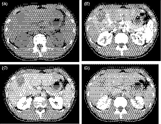
A, Plain computed tomography (CT) revealed multiple tumors with low‐density areas in both hepatic lobes. B‐D, Contrast‐enhanced CT showed the tumor with a slight circular enhancement in the early phase. The enhancement was prolonged to the delayed phase, up to 40 × 46 mm in size
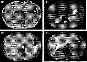
A and B, Magnetic resonance imaging (MRI) showed the tumor with a hypointensity on the T1‐weighted images and a hyperintensity on the T2‐weighted images. C and D, The dynamic MRI study showed the tumor with a heterogeneous enhancement in the early phase and a defect of enhancement in the Kupffer phase
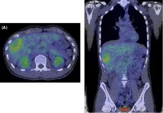
A and B, Fluorin‐18 fluorodeoxyglucose positron emission tomography CT revealed that the tumors had a high accumulation, with a maximum standardized uptake value of 4.9, unlike the other organs
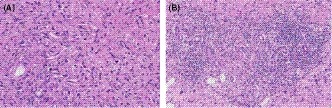
A and B, The histopathological findings revealed that the epithelioid cells were infiltrating the hepatic sinusoids invasively or substitutability. The tumor cells also infiltrated the portal vein and hepatic vein

A and B, Contrast‐enhanced CT conducted 12 mo after liver transplantation showed no finding of recurrence or metastasis in the liver graft or the other organs
References
-
- Weiss SW, Enzinger FM. Epithelioid hemangioendothelioma: a vascular tumor often mistaken for a carcinoma. Cancer. 1982;50:970‐981. - PubMed
-
- Mehrabi A, Kashfi A, Fonouni H, et al. Primary malignant hepatic epithelioid hemangioendothelioma: a comprehensive review of the literature with emphasis on the surgical therapy. Cancer. 2006;107:2108‐2121. - PubMed
-
- Lerut JP, Orlando G, Adam R, et al. for European Liver Transplant Registry. The place of liver transplantation in the treatment of hepatic epitheloid hemangioendothelioma: report of the European Liver Transplant Registry. Ann Surg. 2007;246:949‐957. - PubMed
-
- Sieghart W, Fuereder T, Schmid K, et al. Mammalian target of rapamycin pathway activity in hepatocellular carcinomas of patients undergoing liver transplantation. Transplantation. 2007;83:425‐432. - PubMed
-
- Sahin F, Kannangai R, Adegbola O, Wang J, Su G, Torbenson M. mTOR and P70 S6 kinase expression in primary liver neoplasms. Clin Cancer Res. 2004;10:8421‐8425. - PubMed
Publication types
LinkOut - more resources
Full Text Sources
Research Materials
Miscellaneous
