. 2019 Nov 26:7:296.
doi: 10.3389/fcell.2019.00296. eCollection 2019.

TNFα Induces Müller Glia to Transition From Non-proliferative Gliosis to a Regenerative Response in Mutant Zebrafish Presenting Chronic Photoreceptor Degeneration

Affiliations

TNFα Induces Müller Glia to Transition From Non-proliferative Gliosis to a Regenerative Response in Mutant Zebrafish Presenting Chronic Photoreceptor Degeneration

Maria Iribarne et al. Front Cell Dev Biol. .

Abstract

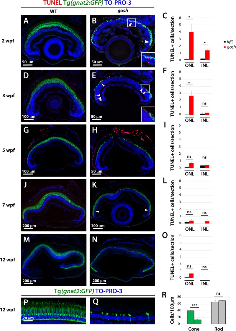
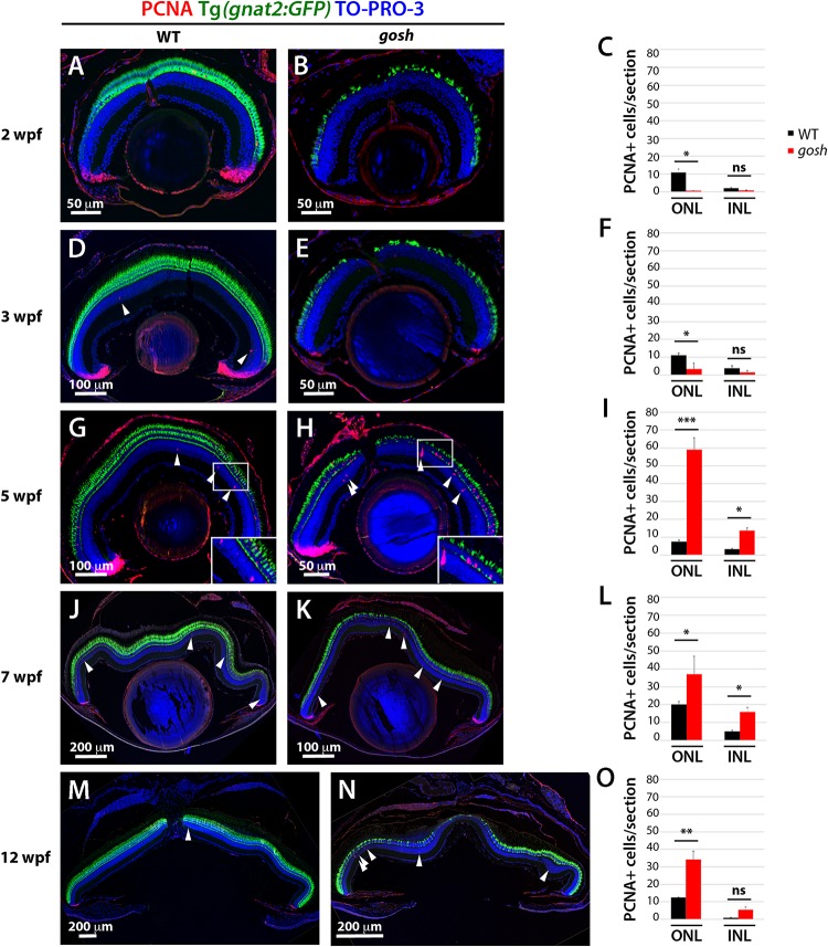
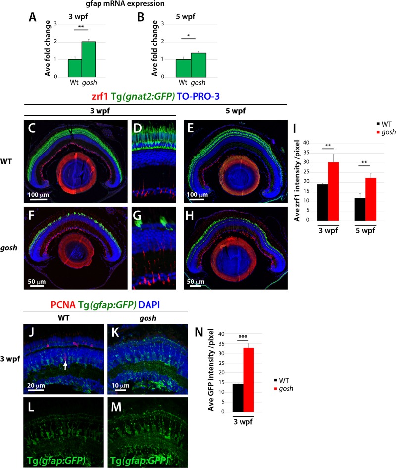
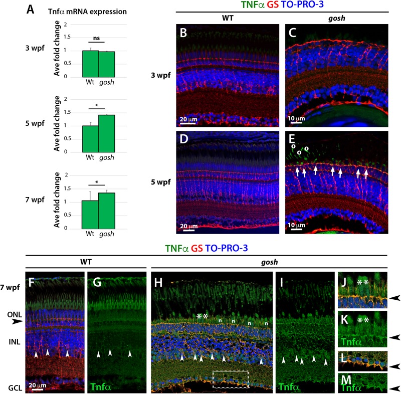
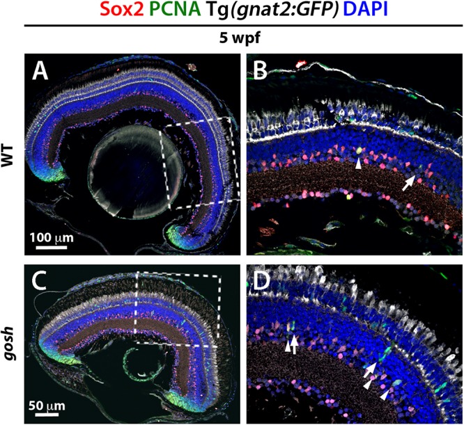
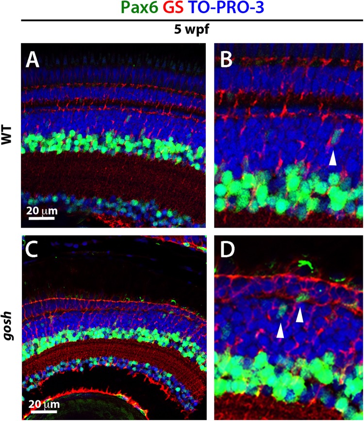
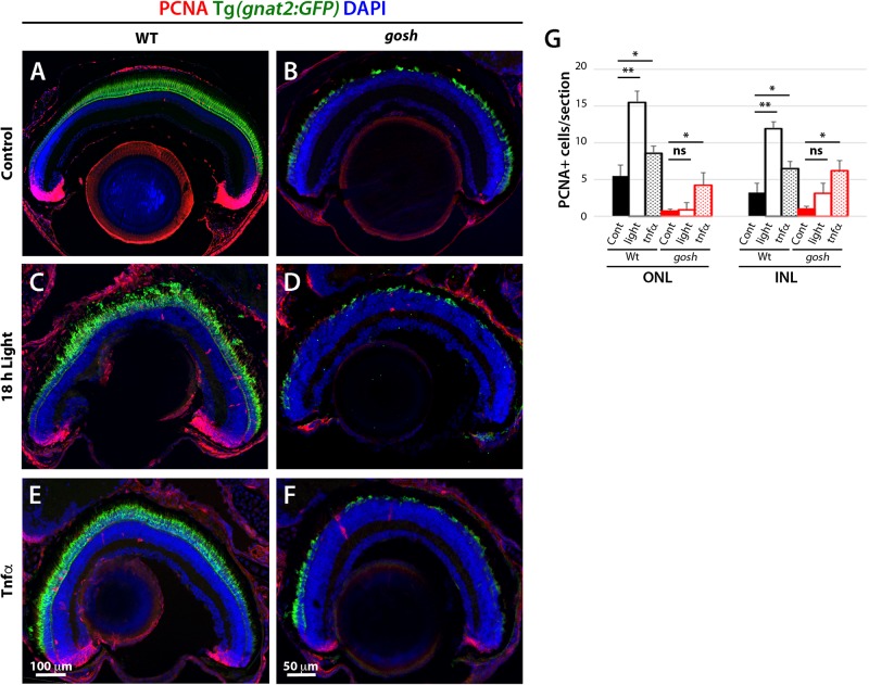
Unlike mammals, zebrafish have the capacity to regenerate neurons in response to damage. Most zebrafish retinal injury models employ acute damage, which is unlike the chronic, gradual damage that occurs in human retinal diseases. Here, we studied the regenerative response in the zebrafish aipl1b mutant, gold rush (gosh). In gosh mutants, both cones and rods degenerate by 3 weeks post-fertilization (wpf). Müller glia do not exhibit a regenerative response by 3 wpf; however, they do present non-proliferative gliosis. Only at 5 wpf, is proliferation of Müller cells and rod precursor cells activated. Rods start to recover at 5 wpf and by 12 wpf they reach a level of recovery comparable to wild type, but cones remain absent in the adult stage. TNFα was detected in degenerating cones at 5-7 wpf and in Müller glia at 7 wpf in gosh mutants. At 5 wpf, proliferating Müller glia express Sox2, followed by Pax6 expression in neuronal progenitor cells (NPCs), confirming that the neuronal regeneration program is activated in gosh mutants after 5 wpf. Although acute light-induced damage did not activate proliferation of Müller glia, TNFα injection caused Müller glia to commence a proliferative response at 3 wpf in gosh mutants. These results suggest that Müller glia transition from non-proliferative gliosis to a regenerative state in gosh mutants, and that ectopic introduction of TNFα promotes this Müller cell transition even at 3 wpf. Thus, zebrafish gosh mutants provide a useful model to investigate mechanisms underlying retinal regeneration in a chronic photoreceptor degeneration model.

Keywords: Aipl1; Müller glia; genetic mutant; photoreceptor degeneration; regeneration; rod precursors; zebrafish.


Figures

FIGURE 1
FIGURE 1
Retinal apoptosis occurs transiently at 2–3 wpf and ceases by 5 wpf in gosh mutants. TUNEL was assessed on retinal slides of wild-type sibling (A,D,G,J,M) and gosh mutant retinas (B,E,H,K,N) at 2, 3, 5, 7, and 12 wpf. Apoptotic cells in the ONL are indicated by arrows. The transgenic line Tg(gnat2:GFP) is used to label cone photoreceptors. Nuclei are counterstained with TO-PRO-3. Wild-type sibling retinas show very low numbers of TUNEL-labeled cells at all stages (A,D,G,J,M). In gosh mutants, numbers of apoptotic cells are higher in the ONL at 2–3 wpf and in the INL at 2 wpf (B,E, insets show TUNEL+ cells in the ONL). Quantification of TUNEL-positive nuclei in the ONL and INL was performed (C,F,I,L,O). Bars and lines indicate means ± SEM, n: 37. Black and red bars: wild-type sibling and gosh mutants. Central retina of control and gosh retinas at 12 wpf show similar rod layer thickness, but cone photoreceptor shows a reduce number of cones (P–R, control left bars, gosh right bars) (ns, p > 0.05; p < 0.05; and ∗∗∗p < 0.001). ONL, outer nuclear layer; INL, inner nuclear layer.
FIGURE 2
FIGURE 2
Proliferation of Müller glia, NPCs, and rod precursors starts after 5 wpf in gosh mutants. Labeling of wild-type sibling (A,D,G,J,M) and gosh mutant retinas (B,E,H,K,N) at 2, 3, 5, 7, and 12 wpf with anti-PCNA antibody. Cones are visualized with the Tg(gnat2:GFP) transgenic line, and nuclei are counterstained with TO-PRO-3. In wild-type retinas, retinal stem and progenitor cells in the CMZ express PCNA, indicating proliferation of retinal progenitor cells. Furthermore, PCNA-positive cells are observed in the ONL and INL, corresponding to rod precursors, and Müller glia/NPCs, respectively, at all stages studied (A,D,G,J,M). In gosh mutants, PCNA expression is absent in the CMZ, as well as in the ONL and INL at 2–3 wpf (B,E). At 5 wpf, PCNA-positive ONL and INL cells are drastically increased in gosh mutants, suggesting that Müller glia and rod precursor cells start cell proliferation (H). Insets show PCNA-positive cells (G,H). Numbers of PCNA-positive cells in the ONL and INL indicate that proliferation becomes maximal at 5 wpf in gosh mutants and maintains higher levels than wild type until 12 wpf (C,F,I,L,O). Bars and lines indicate mean ± SEM, n: 3–9. Black and red bars: wild-type sibling and gosh mutant. Arrowhead depict PCNA-positive cells in the INL (ns, p > 0.05; p < 0.05; ∗∗p < 0.01; and ∗∗∗p < 0.001).
FIGURE 3
FIGURE 3
Non-proliferative gliotic response in gosh mutants at 3 wpf. (A,B) Upregulation of GFAP mRNA expression in gosh samples at 3 and 5 wpf compared with control samples. (C–H) Paraffin sections labeled with zrf1 antibody, which recognizes GFAP. Cones are visualized with GFP expression of T(gnat2:GFP). Nuclei are stained with TO-PRO-3. In wild type, inner radial processes of Müller glia are faintly stained with zrf1 antibody at 3 and 5 wpf (C–E). However, in gosh mutants, radial processes of Müller glia are more intensely labeled at 3 and 5 wpf, indicating cell hypertrophy with upregulation of GFAP (F–H). (I) Histogram of GFAP-positive area in wild-type and gosh mutant retinas at 3 and 5 wpf. GFAP signals are higher in gosh mutants than in wild type at both 3 and 5 wpf. (J–M) Tg(gfap:GFP)nt11 visualizes Müller glia at 3 wpf. Proliferative Müller glia, NPCs, and rod precursor cells are labeled with PCNA antibody, and nuclei are counterstained with DAPI. In wild-type retinas, GFAP is observed in cell bodies and apico-basal extended processes of Müller glia (J,L). Some Müller glia express PCNA (J, arrow). PCNA-positive cells are also observed in the ONL, indicating persistent neurogenesis to produce rod photoreceptors. In contrast, PCNA expression is absent or very low in gosh mutants at 3 wpf (K). GFAP is upregulated in Müller cells, which show a greater number of cell processes (K,M). Notice the strong GFP fluorescence in the ONL, where photoreceptors are degenerating. (N) Histogram of gfap-positive area in control and gosh retinas depicts the increase of fluorescence in the mutant retina.
FIGURE 4
FIGURE 4
TNFα is detected in cones at 5 wpf and in Müller glia at 7 wpf in gosh mutants. (A) Quantitative PCR histogram of 3, 5, and 7 wpf of control and gosh samples. 3 wpf of control and gosh samples displayed similar levels of TNFα mRNA. Upregulation of TNFα mRNA at 5 and 7 wpf in gosh samples relative to control samples. (B–M) Paraffin sections of wild-type sibling and gosh mutant retinas were labeled with antibodies against TNFα and GS at 3, 5, and 7 wpf. Müller glia are visualized as cells with nuclei located in the INL and radial processes that span the apico-basal axis of the neural retina. Wild-type sibling retinas exhibit undetectable levels of TNFα (B,D). gosh mutant retinas show no TNFα immunoreactivity at 3 wpf, as in wild-type retinas (C); however, degenerating cones display labeling against TNFα at 5 wpf (E, arrows). Circles in (E) denote autofluorescence. Nuclei of Müller glia are localized in the INL (white arrowhead) and their basal and apical processes reach the basal region of the retinal ganglion cell layer and the outer limiting membrane (black arrowhead) in the ONL in 7-wpf wild-type retinas (F,G). TNFα immunoreactivity is absent, although background signals are observed in the cone outer segment. However, in gosh mutant retina, strong signals are detected in dying cones (H, asterisks) and weaker signal in Müller glia (white arrowhead) and their cell process (H,I). (J,K) indicate high magnification images of the ONL shown in (H, asteriks). (L,M) indicate high magnification images of basal foot shown in (H, dotted box). The outer limiting membrane (J,K, black arrowhead) and the basal feet of Müller glia (L,M) are strongly stained. Amacrine and ganglion cells show faint TNFα-positive signal (H,I). n: nuclei of cone photoreceptors (ns, p > 0.05; p < 0.05).
FIGURE 5
FIGURE 5
Müller glia express Sox2 during the regeneration response in gosh mutants. Wild-type and gosh mutant retinas at 5 wpf labeled with anti-Sox2 and anti-PCNA antibodies, and DAPI. Tg(gnat2:GFP) transgene visualizes cones. Wild-type retinas display Sox2 expression in the CMZ and amacrine cells (A,C, dotted boxes show the area magnifies in B,D). Müller glia exhibit faint expression of Sox2. At 5 wpf, gosh mutant retinas show that Sox2 expression is colocalized with PCNA in the INL (D, arrowheads) probably in Müller glia, whereas subsequently differentiating NPCs express only PCNA (D, arrows).
FIGURE 6
FIGURE 6
NPCs express Pax6 during the regenerative response in gosh mutants. Wild-type and gosh mutant retinas labeled with antibodies against Pax6 and GS, which are expressed in NPCs and Müller glia, respectively. In wild-type retinas, Pax6 is observed in amacrine cells and retinal ganglion cells (A,B). Weaker signals were observed in NPCs migrating to the ONL (B, arrowhead). Five-wpf gosh mutant retinas show that Pax6 is expressed in amacrine cells and retinal ganglion cells, as well as probable NPCs migrating toward the ONL (C,D, arrowheads).
FIGURE 7
FIGURE 7
Proliferation of Müller glia and rod precursor cells is not activated by light-induced damage, but by TNFα in gosh mutants at 3 wpf. Wild-type and gosh mutant retinas at 3 wpf are labeled with anti-PCNA antibody. Nuclei are stained with DAPI. The transgene Tg(gnat2:GFP) visualizes cone photoreceptors. In wild-type, undamaged controls, PCNA is expressed in the CMZ, and PCNA-positive cells are observed in the INL and ONL (A). However, the PCNA signal is almost absent in gosh mutant retinas (B). Treatment for 18 h under intense light damages photoreceptors (C,D). Wild-type sibling retinas showed an increase in the number of PCNA-positive cells located in the INL and ONL. On the other hand, the PCNA signal is still absent in gosh mutant retinas. TNFα intravitreal injections increase the number of PCNA-positive cells in wild-type sibling and gosh mutant retinas (E,F). Histogram depicting the number of PCNA-positive cells (G). Bars and lines indicate mean ± SEM, n: 611. Black and red bars, wild-type siblings and gosh mutants. Filled bars, no treatment; Open bars, photodamaged samples; Dotted bars, TNFα-injected sample (ns, p > 0.05; p < 0.05; ∗∗p < 0.01).

Similar articles

Cited by

References

    1. Bernardos R. L., Barthel L. K., Meyers J. R., Raymond P. A. (2007). Late-stage neuronal progenitors in the retina are radial Muller glia that function as retinal stem cells. J. Neurosci. 27 7028–7040. 10.1523/JNEUROSCI.1624-07.2007 - DOI - PMC - PubMed
    1. Bringmann A., Wiedemann P. (2012). Muller glial cells in retinal disease. Ophthalmologica 227 1–19. 10.1159/000328979 - DOI - PubMed
    1. Campbell L. J., Hyde D. R. (2017). Opportunities for CRISPR/Cas9 gene editing in retinal regeneration research. Front. Cell Dev. Biol. 5:99. 10.3389/fcell.2017.00099 - DOI - PMC - PubMed
    1. Conner C., Ackerman K. M., Lahne M., Hobgood J. S., Hyde D. R. (2014). Repressing notch signaling and expressing TNFα are sufficient to mimic retinal regeneration by inducing Muller glial proliferation to generate committed progenitor cells. J. Neurosci. 34 14403–14419. 10.1523/JNEUROSCI.0498-14.2014 - DOI - PMC - PubMed
    1. Craig S. E., Calinescu A. A., Hitchcock P. F. (2008). Identification of the molecular signatures integral to regenerating photoreceptors in the retina of the zebra fish. J. Ocul. Biol. Dis. Infor. 1 73–84. 10.1007/s12177-008-9011-5 - DOI - PMC - PubMed

LinkOut - more resources