

A machine learning cardiac magnetic resonance approach to extract disease features and automate pulmonary arterial hypertension diagnosis
- PMID: 31998956
- PMCID: PMC7822638
- DOI: 10.1093/ehjci/jeaa001
A machine learning cardiac magnetic resonance approach to extract disease features and automate pulmonary arterial hypertension diagnosis
Abstract
Aims: Pulmonary arterial hypertension (PAH) is a progressive condition with high mortality. Quantitative cardiovascular magnetic resonance (CMR) imaging metrics in PAH target individual cardiac structures and have diagnostic and prognostic utility but are challenging to acquire. The primary aim of this study was to develop and test a tensor-based machine learning approach to holistically identify diagnostic features in PAH using CMR, and secondarily, visualize and interpret key discriminative features associated with PAH.
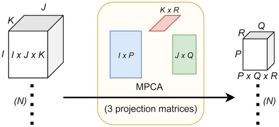
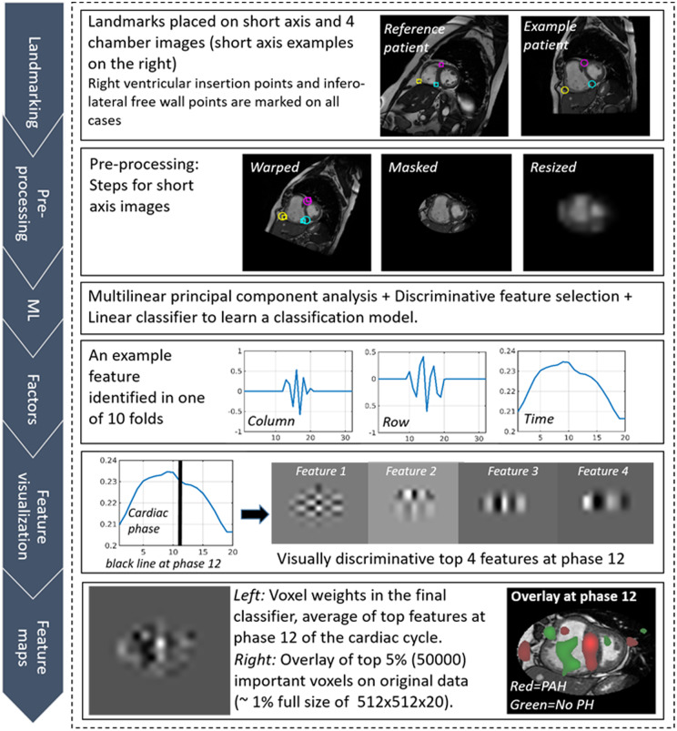
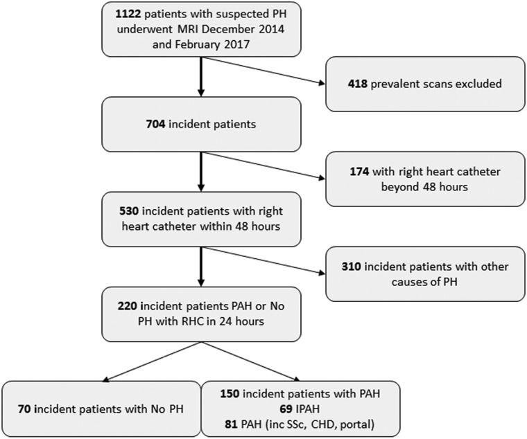
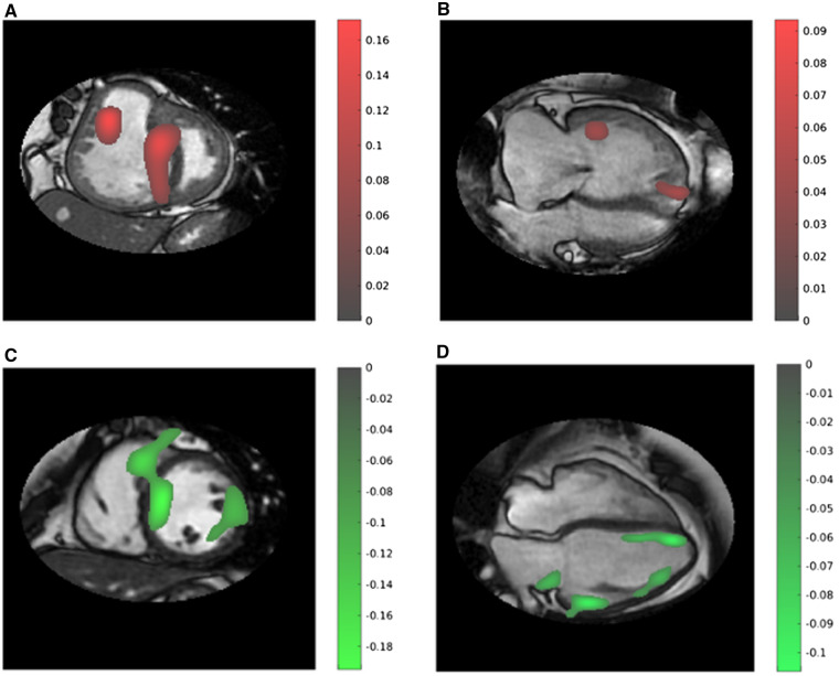
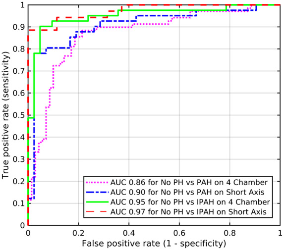
Methods and results: Consecutive treatment naive patients with PAH or no evidence of pulmonary hypertension (PH), undergoing CMR and right heart catheterization within 48 h, were identified from the ASPIRE registry. A tensor-based machine learning approach, multilinear subspace learning, was developed and the diagnostic accuracy of this approach was compared with standard CMR measurements. Two hundred and twenty patients were identified: 150 with PAH and 70 with no PH. The diagnostic accuracy of the approach was high as assessed by area under the curve at receiver operating characteristic analysis (P < 0.001): 0.92 for PAH, slightly higher than standard CMR metrics. Moreover, establishing the diagnosis using the approach was less time-consuming, being achieved within 10 s. Learnt features were visualized in feature maps with correspondence to cardiac phases, confirming known and also identifying potentially new diagnostic features in PAH.
Conclusion: A tensor-based machine learning approach has been developed and applied to CMR. High diagnostic accuracy has been shown for PAH diagnosis and new learnt features were visualized with diagnostic potential.
Keywords: diagnosis; machine learning; pulmonary arterial hypertension; right ventricle; tensor.
© The Author(s) 2020. Published by Oxford University Press on behalf of the European Society of Cardiology.
Figures
References
-
- Kiely DG, Elliot CA, Sabroe I, Condliffe R.. Pulmonary hypertension: diagnosis and management. BMJ 2013;346:f2028. - PubMed
-
- Swift AJ, Rajaram S, Condliffe R, Capener D, Hurdman J, Elliot C. et al. Pulmonary artery relative area change detects mild elevations in pulmonary vascular resistance and predicts adverse outcome in pulmonary hypertension. Invest Radiol 2012;47:571–7. - PubMed
-
- Schäfer M, Myers C, Brown RD, Frid MG, Tan W, Hunter K. et al. Pulmonary arterial stiffness: toward a new paradigm in pulmonary arterial hypertension pathophysiology and assessment. Curr Hypertens Rep 2016;18:4.. - PubMed
-
- Gan C-J, Lankhaar J-W, Westerhof N, Marcus JT, Becker A, Twisk JWR. et al. Noninvasively assessed pulmonary artery stiffness predicts mortality in pulmonary arterial hypertension. Chest 2007;132:1906–12. - PubMed
