

Top-Down Characterization of Denatured Proteins and Native Protein Complexes Using Electron Capture Dissociation Implemented within a Modified Ion Mobility-Mass Spectrometer
- PMID: 31999103
- PMCID: PMC7847745
- DOI: 10.1021/acs.analchem.9b04763
Top-Down Characterization of Denatured Proteins and Native Protein Complexes Using Electron Capture Dissociation Implemented within a Modified Ion Mobility-Mass Spectrometer
Abstract
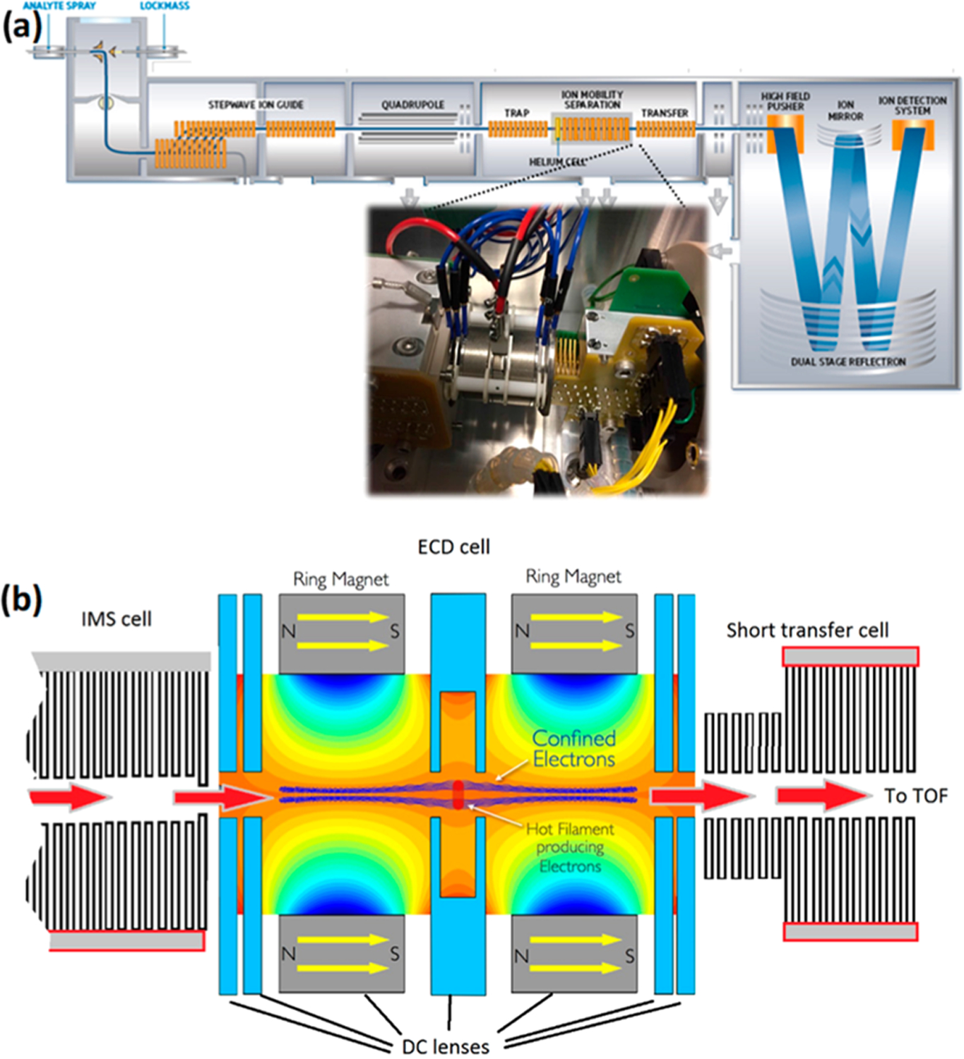
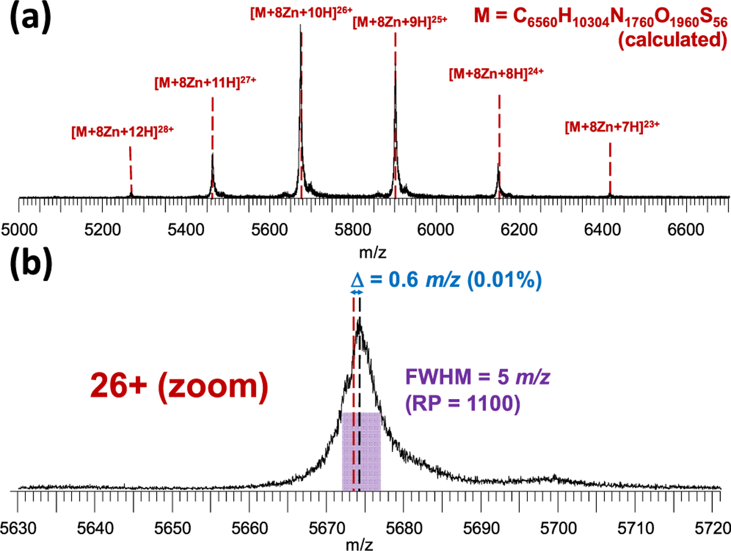
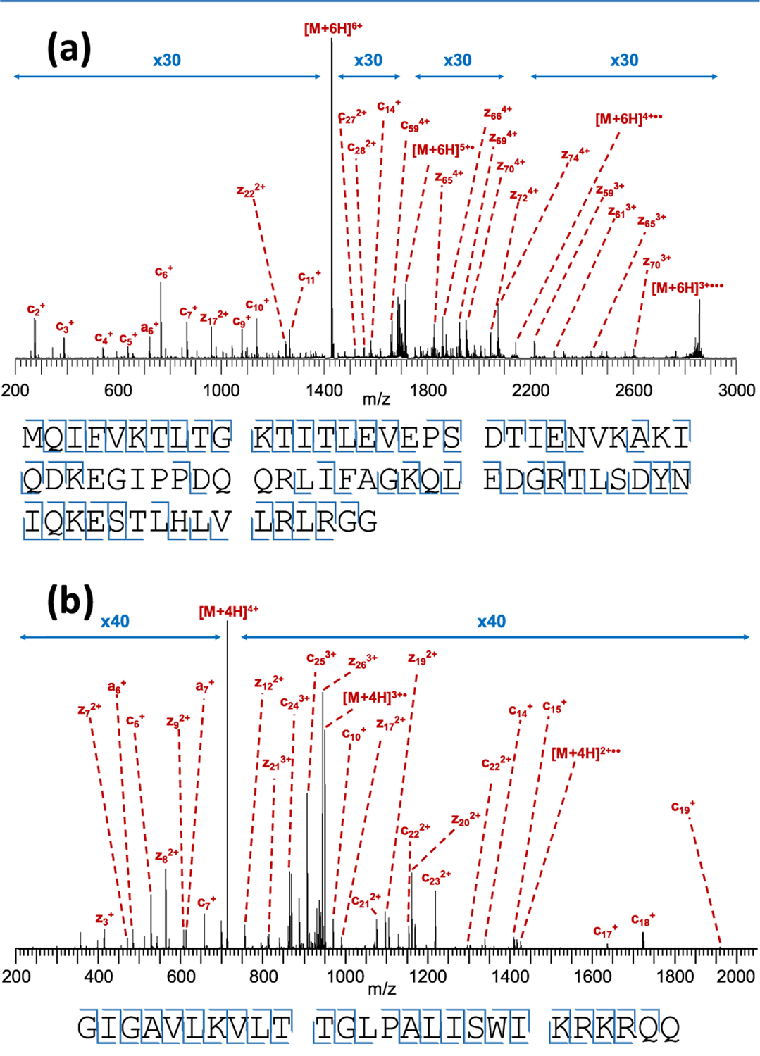
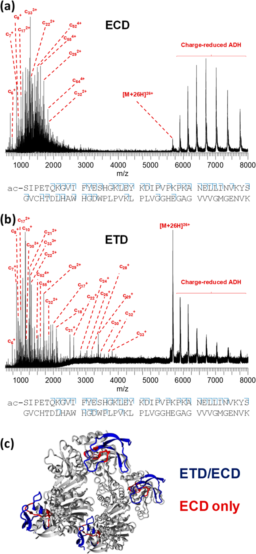
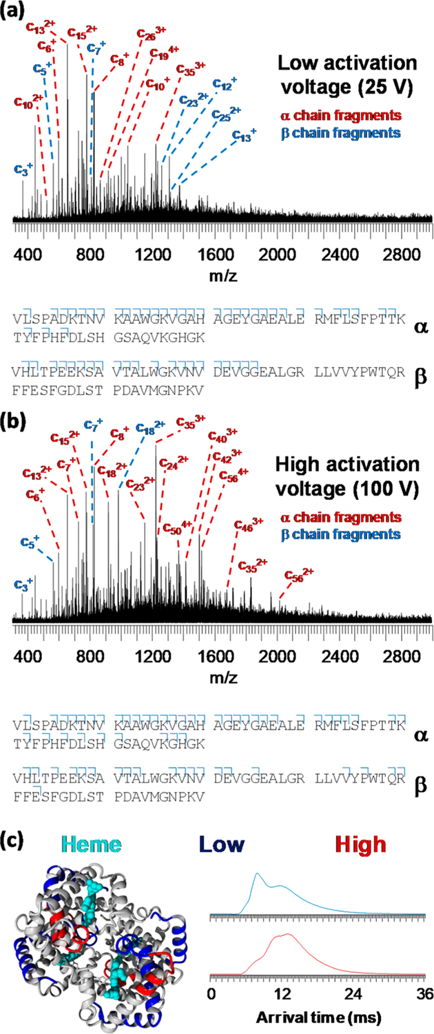
Electron-based fragmentation methods have revolutionized biomolecular mass spectrometry, in particular native and top-down protein analysis. Here, we report the use of a new electromagnetostatic cell to perform electron capture dissociation (ECD) within a quadrupole/ion mobility/time-of-flight mass spectrometer. This cell was installed between the ion mobility and time-of-flight regions of the instrument, and fragmentation was fast enough to be compatible with mobility separation. The instrument was already fitted with electron transfer dissociation (ETD) between the quadrupole and mobility regions prior to modification. We show excellent fragmentation efficiency for denatured peptides and proteins without the need to trap ions in the gas phase. Additionally, we demonstrate native top-down backbone fragmentation of noncovalent protein complexes, leading to comparable sequence coverage to what was achieved using the instrument's existing ETD capabilities. Limited collisional ion activation of the hemoglobin tetramer before ECD was reflected in the observed fragmentation pattern, and complementary ion mobility measurements prior to ECD provided orthogonal evidence of monomer unfolding within this complex. The approach demonstrated here provides a powerful platform for both top-down proteomics and mass spectrometry-based structural biology studies.
Conflict of interest statement
The authors declare no competing financial interest.
Figures
Similar articles
-
Capillary Zone Electrophoresis-Electron-Capture Collision-Induced Dissociation on a Quadrupole Time-of-Flight Mass Spectrometer for Top-Down Characterization of Intact Proteins.J Am Soc Mass Spectrom. 2021 Jun 2;32(6):1361-1369. doi: 10.1021/jasms.0c00484. Epub 2021 Mar 22. J Am Soc Mass Spectrom. 2021. PMID: 33749270 Free PMC article.
-
Electron transfer dissociation provides higher-order structural information of native and partially unfolded protein complexes.Proteomics. 2015 Aug;15(16):2813-22. doi: 10.1002/pmic.201400516. Epub 2015 Jul 22. Proteomics. 2015. PMID: 26081219
-
Characterization of top-down ETD in a travelling-wave ion guide.Methods. 2015 Nov 1;89:22-9. doi: 10.1016/j.ymeth.2015.05.019. Epub 2015 May 23. Methods. 2015. PMID: 26014039
-
Radical solutions: Principles and application of electron-based dissociation in mass spectrometry-based analysis of protein structure.Mass Spectrom Rev. 2018 Nov;37(6):750-771. doi: 10.1002/mas.21560. Epub 2018 Feb 9. Mass Spectrom Rev. 2018. PMID: 29425406 Free PMC article. Review.
-
'Top down' protein characterization via tandem mass spectrometry.J Mass Spectrom. 2002 Jul;37(7):663-75. doi: 10.1002/jms.346. J Mass Spectrom. 2002. PMID: 12124999 Review.
Cited by
-
Gas-Phase Ion/Ion Chemistry for Structurally Sensitive Probes of Gaseous Protein Ion Structure: Electrostatic and Electrostatic to Covalent Cross-Linking.Int J Mass Spectrom. 2021 May;463:116549. doi: 10.1016/j.ijms.2021.116549. Epub 2021 Feb 17. Int J Mass Spectrom. 2021. PMID: 33716558 Free PMC article.
-
Characterization of an Omnitrap-Orbitrap Platform Equipped with Infrared Multiphoton Dissociation, Ultraviolet Photodissociation, and Electron Capture Dissociation for the Analysis of Peptides and Proteins.Anal Chem. 2023 Aug 15;95(32):12039-12046. doi: 10.1021/acs.analchem.3c01899. Epub 2023 Aug 3. Anal Chem. 2023. PMID: 37534599 Free PMC article.
-
Capillary Zone Electrophoresis-Electron-Capture Collision-Induced Dissociation on a Quadrupole Time-of-Flight Mass Spectrometer for Top-Down Characterization of Intact Proteins.J Am Soc Mass Spectrom. 2021 Jun 2;32(6):1361-1369. doi: 10.1021/jasms.0c00484. Epub 2021 Mar 22. J Am Soc Mass Spectrom. 2021. PMID: 33749270 Free PMC article.
-
Revealing the Fates of Proteins in the Gas Phase.Int J Mass Spectrom. 2024 Oct;504:117312. doi: 10.1016/j.ijms.2024.117312. Epub 2024 Jul 30. Int J Mass Spectrom. 2024. PMID: 39184132
-
Tandem surface-induced dissociation of protein complexes on an ultrahigh resolution platform.Int J Mass Spectrom. 2021 Mar;461:116503. doi: 10.1016/j.ijms.2020.116503. Epub 2021 Jan 5. Int J Mass Spectrom. 2021. PMID: 33889055 Free PMC article.
References
-
- Schadt EE; Turner S; Kasarskis A Hum. Mol. Genet 2010, 19, R227–240. - PubMed
Publication types
MeSH terms
Substances
Grants and funding
LinkOut - more resources
Full Text Sources
