

CT-pathologic correlation of non-calcified atherosclerotic arterial plaques: a study using carotid endarterectomy specimens
- PMID: 31999208
- PMCID: PMC7217582
- DOI: 10.1259/bjr.20190901
CT-pathologic correlation of non-calcified atherosclerotic arterial plaques: a study using carotid endarterectomy specimens
Abstract
Objective: Pathologic features of atherosclerotic plaques on CT are not established. We compared CT values among pathologically confirmed plaque constituents and evaluated their ability to distinguish plaque constituents.
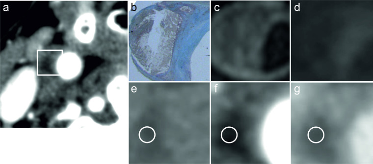
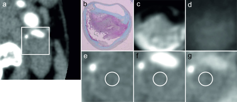
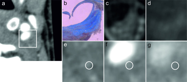
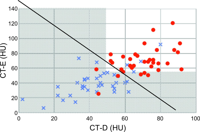
Methods: 50 histopathological images of carotid endarterectomy samples from 10 males and 2 females (age 54-74 years, average 65.9 years) were examined. We compared pre-operative CT [pre-contrast (CT-P), early post-contrast phase (CT-E), delayed post-contrast phase (CT-D)] of lipid-rich necrotic core (NC) and fibrous tissue (F) plaque components with pathological images. The ability of features to differentiate plaque components using several discrimination techniques were compared.
Results: CT values of NC and F were 36 ± 13, 45 ± 11 (mean ± standard deviation, Hounsfield unit, HU), 41 ± 17, 69 ± 18, and 44 ± 16, 70 ± 13 in CT-P (p < 0.01), CT-E (p < 0.0001), and CT-D (p < 0.0001), respectively. The threshold, sensitivity, and accuracy for distinguishing NC from F were 44 HU, 74%, and 68%; 55 HU, 85%, and 85%; and 63 HU, 92%, and 84% in CTP, CT-E, and CT-D, respectively. CT-P had lower accuracy than CT-E and CT-D (both p < 0.05), but CT-E and CT-D were similar. CT-E and CT-D yielded 90 and 91% sensitivity and accuracy, respectively in linear discrimination analysis.
Conclusion: In both pre- and post-contrast CT, CT values were lower in NC than F. Although values overlapped, using two-phase post-contrast CTs improved discrimination ability.
Advances in knowledge: Our findings may help to establish computer-aided diagnosis of vulnerable atherosclerotic plaques in future.
Figures
Similar articles
-
Identification of vulnerable carotid plaque with histologically validated CT-derived plaque maps.Br J Radiol. 2023 Jul;96(1147):20220982. doi: 10.1259/bjr.20220982. Epub 2023 May 15. Br J Radiol. 2023. PMID: 37183910 Free PMC article.
-
CT Attenuation Analysis of Carotid Intraplaque Hemorrhage.AJNR Am J Neuroradiol. 2018 Jan;39(1):131-137. doi: 10.3174/ajnr.A5461. Epub 2017 Nov 30. AJNR Am J Neuroradiol. 2018. PMID: 29191874 Free PMC article.
-
Assessment of carotid plaque stability based on the dynamic enhancement pattern in plaque components with multidetector CT angiography.Stroke. 2012 Feb;43(2):393-8. doi: 10.1161/STROKEAHA.111.635953. Epub 2011 Nov 17. Stroke. 2012. PMID: 22096033
-
Contemporary carotid imaging: from degree of stenosis to plaque vulnerability.J Neurosurg. 2016 Jan;124(1):27-42. doi: 10.3171/2015.1.JNS142452. Epub 2015 Jul 31. J Neurosurg. 2016. PMID: 26230478 Review.
-
Molecular imaging of carotid plaque vulnerability.Cerebrovasc Dis. 2015;39(1):5-12. doi: 10.1159/000369123. Epub 2014 Dec 24. Cerebrovasc Dis. 2015. PMID: 25547782 Review.
Cited by
-
Carotid plaque characteristics by computed Tomography: A diagnostic accuracy systematic review.Int J Cardiol Heart Vasc. 2025 Mar 21;58:101656. doi: 10.1016/j.ijcha.2025.101656. eCollection 2025 Jun. Int J Cardiol Heart Vasc. 2025. PMID: 40213415 Free PMC article. Review.
-
Utility of vector flow mapping technology in quantitative assessment of carotid wall shear stress in hypertensive patients: A preliminary study.Front Cardiovasc Med. 2022 Oct 28;9:967763. doi: 10.3389/fcvm.2022.967763. eCollection 2022. Front Cardiovasc Med. 2022. PMID: 36386366 Free PMC article.
References
-
- Global Health Observatory (GHO) data World health Organization web site.. Available from: https://www.who.int/gho/mortality_burden_disease/en/ [Accessed 17thJuly, 2019].
-
- Little WC, Constantinescu M, Applegate RJ, Kutcher MA, Burrows MT, Kahl FR, et al. . Can coronary angiography predict the site of a subsequent myocardial infarction in patients with mild-to-moderate coronary artery disease? Circulation 1988; 78(5 Pt 1): 1157–66. doi: 10.1161/01.CIR.78.5.1157 - DOI - PubMed
-
- North American symptomatic carotid endarterectomy trial Collaborators. beneficial effect of carotid endarterectomy in symptomatic patients with high-grade carotid stenosis. N Engl J Med 1991; 325: 445–53. - PubMed
Publication types
MeSH terms
Substances
LinkOut - more resources
Full Text Sources
Research Materials
