

The Role of the Epicardium During Heart Development and Repair
- PMID: 31999538
- PMCID: PMC7000171
- DOI: 10.1161/CIRCRESAHA.119.315857
The Role of the Epicardium During Heart Development and Repair
Abstract
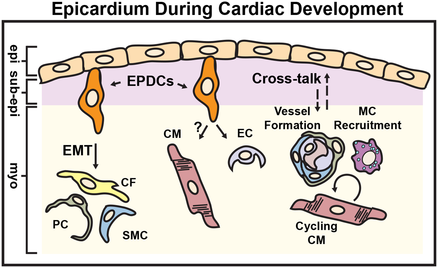
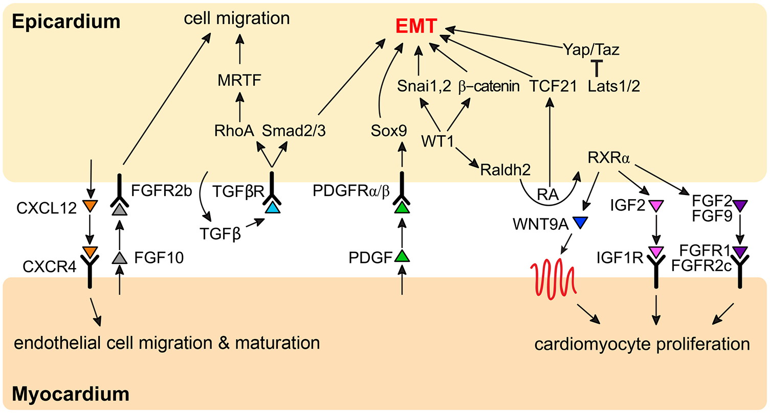
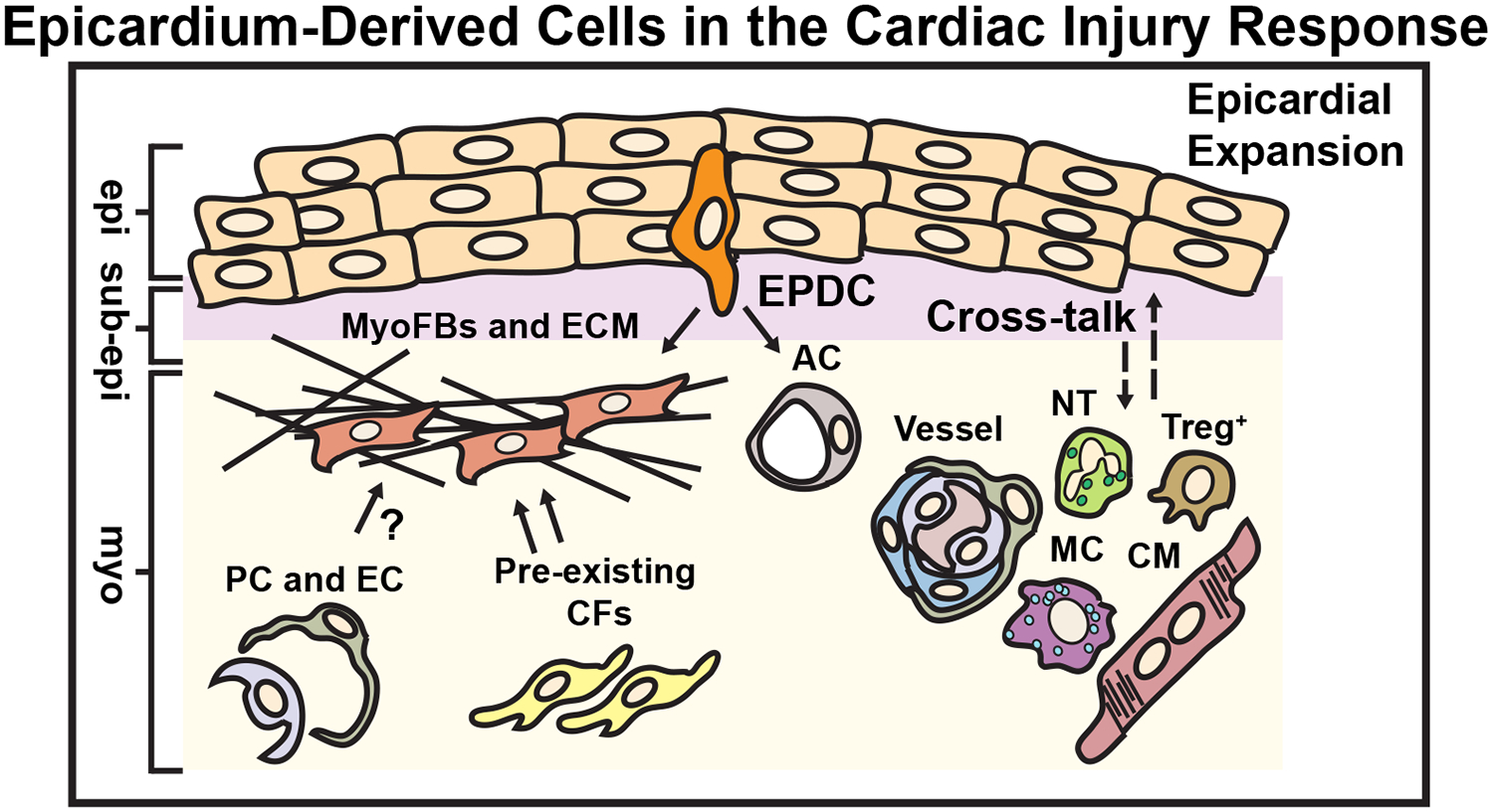
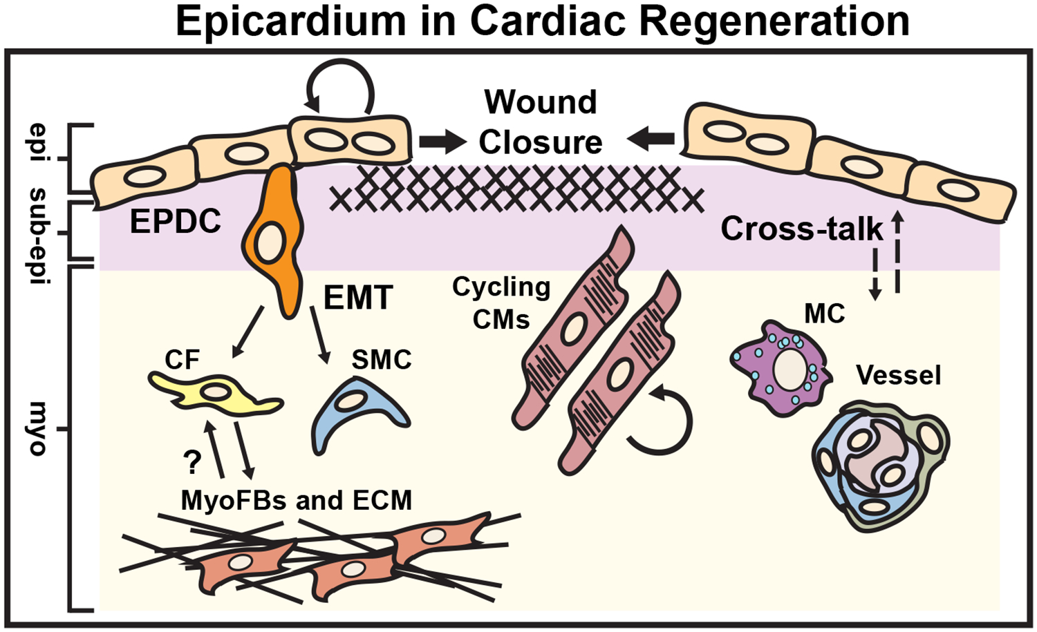
The heart is lined by a single layer of mesothelial cells called the epicardium that provides important cellular contributions for embryonic heart formation. The epicardium harbors a population of progenitor cells that undergo epithelial-to-mesenchymal transition displaying characteristic conversion of planar epithelial cells into multipolar and invasive mesenchymal cells before differentiating into nonmyocyte cardiac lineages, such as vascular smooth muscle cells, pericytes, and fibroblasts. The epicardium is also a source of paracrine cues that are essential for fetal cardiac growth, coronary vessel patterning, and regenerative heart repair. Although the epicardium becomes dormant after birth, cardiac injury reactivates developmental gene programs that stimulate epithelial-to-mesenchymal transition; however, it is not clear how the epicardium contributes to disease progression or repair in the adult. In this review, we will summarize the molecular mechanisms that control epicardium-derived progenitor cell migration, and the functional contributions of the epicardium to heart formation and cardiomyopathy. Future perspectives will be presented to highlight emerging therapeutic strategies aimed at harnessing the regenerative potential of the fetal epicardium for cardiac repair.
Keywords: fibrosis; growth and development; myocardial ischemia; paracrine communication; regeneration.
Figures
Similar articles
-
The epicardium in cardiac repair: from the stem cell view.Pharmacol Ther. 2011 Jan;129(1):82-96. doi: 10.1016/j.pharmthera.2010.09.002. Epub 2010 Oct 19. Pharmacol Ther. 2011. PMID: 20937304 Review.
-
The epicardium as a source of multipotent adult cardiac progenitor cells: Their origin, role and fate.Pharmacol Res. 2018 Jan;127:129-140. doi: 10.1016/j.phrs.2017.07.020. Epub 2017 Jul 24. Pharmacol Res. 2018. PMID: 28751220 Review.
-
The epicardium signals the way towards heart regeneration.Stem Cell Res. 2014 Nov;13(3 Pt B):683-92. doi: 10.1016/j.scr.2014.04.007. Epub 2014 Apr 29. Stem Cell Res. 2014. PMID: 24933704 Free PMC article. Review.
-
Epicardium-derived cells in cardiogenesis and cardiac regeneration.Cell Mol Life Sci. 2007 Mar;64(6):692-703. doi: 10.1007/s00018-007-6522-3. Cell Mol Life Sci. 2007. PMID: 17380310 Free PMC article. Review.
-
The epicardium as a hub for heart regeneration.Nat Rev Cardiol. 2018 Oct;15(10):631-647. doi: 10.1038/s41569-018-0046-4. Nat Rev Cardiol. 2018. PMID: 29950578 Free PMC article. Review.
Cited by
-
Single-Cell Transcriptomics of Engineered Cardiac Tissues From Patient-Specific Induced Pluripotent Stem Cell-Derived Cardiomyocytes Reveals Abnormal Developmental Trajectory and Intrinsic Contractile Defects in Hypoplastic Right Heart Syndrome.J Am Heart Assoc. 2020 Oct 20;9(20):e016528. doi: 10.1161/JAHA.120.016528. Epub 2020 Oct 16. J Am Heart Assoc. 2020. PMID: 33059525 Free PMC article.
-
Role of PDGF-A/B Ligands in Cardiac Repair After Myocardial Infarction.Front Cell Dev Biol. 2021 Aug 25;9:669188. doi: 10.3389/fcell.2021.669188. eCollection 2021. Front Cell Dev Biol. 2021. PMID: 34513823 Free PMC article. Review.
-
Exploring the role of epicardial adipose-tissue-derived extracellular vesicles in cardiovascular diseases.iScience. 2024 Feb 29;27(4):109359. doi: 10.1016/j.isci.2024.109359. eCollection 2024 Apr 19. iScience. 2024. PMID: 38510143 Free PMC article. Review.
-
Recapitulating porcine cardiac development in vitro: from expanded potential stem cell to embryo culture models.Front Cell Dev Biol. 2023 May 15;11:1111684. doi: 10.3389/fcell.2023.1111684. eCollection 2023. Front Cell Dev Biol. 2023. PMID: 37261075 Free PMC article.
-
Multiscale drug screening for cardiac fibrosis identifies MD2 as a therapeutic target.Cell. 2024 Dec 12;187(25):7143-7163.e22. doi: 10.1016/j.cell.2024.09.034. Epub 2024 Oct 15. Cell. 2024. PMID: 39413786
References
-
- Benjamin EJ, Muntner P, Alonso A, Bittencourt MS, Callaway CW, Carson AP, Chamberlain AM, Chang AR, Cheng S, Das SR, Delling FN, Djousse L, Elkind MSV, Ferguson JF, Fornage M, Jordan LC, Khan SS, Kissela BM, Knutson KL, Kwan TW, Lackland DT, Lewis TT, Lichtman JH, Longenecker CT, Loop MS, Lutsey PL, Martin SS, Matsushita K, Moran AE, Mussolino ME, O’Flaherty M, Pandey A, Perak AM, Rosamond WD, Roth GA, Sampson UKA, Satou GM, Schroeder EB, Shah SH, Spartano NL, Stokes A, Tirschwell DL, Tsao CW, Turakhia MP, VanWagner LB, Wilkins JT, Wong SS, Virani SS, American Heart Association Council on E, Prevention Statistics C and Stroke Statistics S. Heart Disease and Stroke Statistics-2019 Update: A Report From the American Heart Association. Circulation. 2019;139:e56–e528. - PubMed
-
- Forte E, Furtado MB and Rosenthal N. The interstitium in cardiac repair: role of the immune-stromal cell interplay. Nat Rev Cardiol. 2018;15:601–616. - PubMed
Publication types
MeSH terms
Grants and funding
LinkOut - more resources
Full Text Sources
Other Literature Sources
Medical
