

Recent advances in lab-on-a-chip technologies for viral diagnosis
- PMID: 31999560
- PMCID: PMC7126858
- DOI: 10.1016/j.bios.2020.112041
Recent advances in lab-on-a-chip technologies for viral diagnosis
Abstract
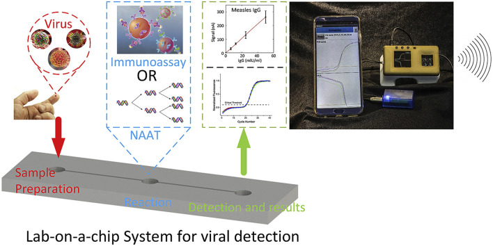
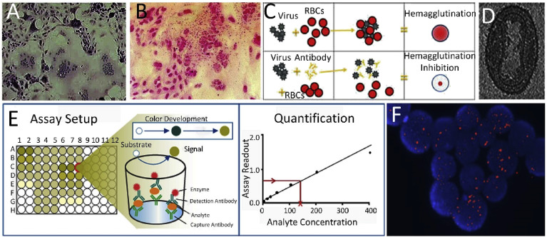
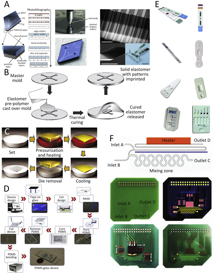
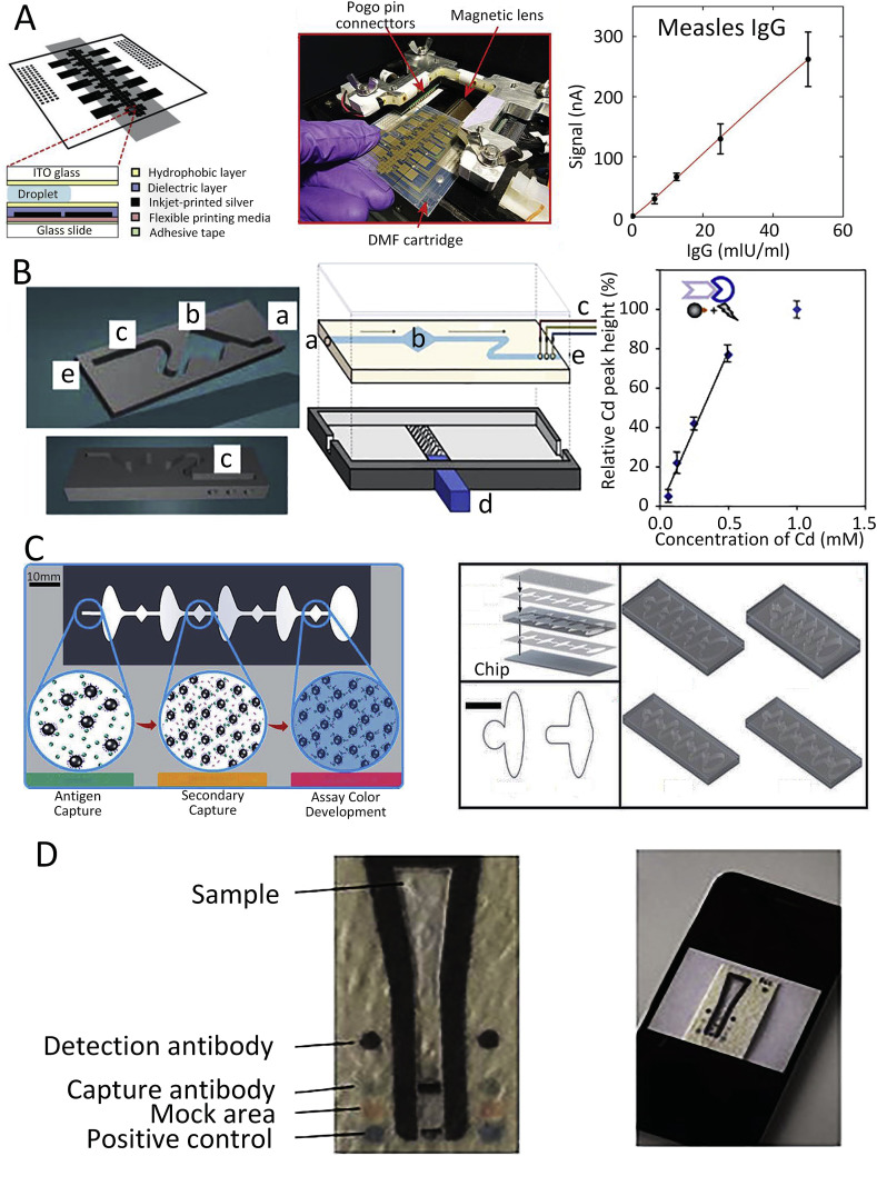
The global risk of viral disease outbreaks emphasizes the need for rapid, accurate, and sensitive detection techniques to speed up diagnostics allowing early intervention. An emerging field of microfluidics also known as the lab-on-a-chip (LOC) or micro total analysis system includes a wide range of diagnostic devices. This review briefly covers both conventional and microfluidics-based techniques for rapid viral detection. We first describe conventional detection methods such as cell culturing, immunofluorescence or enzyme-linked immunosorbent assay (ELISA), or reverse transcription polymerase chain reaction (RT-PCR). These methods often have limited speed, sensitivity, or specificity and are performed with typically bulky equipment. Here, we discuss some of the LOC technologies that can overcome these demerits, highlighting the latest advances in LOC devices for viral disease diagnosis. We also discuss the fabrication of LOC systems to produce devices for performing either individual steps or virus detection in samples with the sample to answer method. The complete system consists of sample preparation, and ELISA and RT-PCR for viral-antibody and nucleic acid detection, respectively. Finally, we formulate our opinions on these areas for the future development of LOC systems for viral diagnostics.
Keywords: Commercialization; Immunoassays; LOC; Microfluidic; Nucleic acid amplification; Viral detection.
Copyright © 2020 Elsevier B.V. All rights reserved.
Conflict of interest statement
Declaration of competing interest The authors declare that they have no known competing financial interests or personal relationships that could have appeared to influence the work reported in this paper.
Figures
Similar articles
-
Integrated sample-to-detection chip for nucleic acid test assays.Biomed Microdevices. 2016 Jun;18(3):44. doi: 10.1007/s10544-016-0069-8. Biomed Microdevices. 2016. PMID: 27165104
-
Highly-integrated lab-on-chip system for point-of-care multiparameter analysis.Lab Chip. 2012 Feb 7;12(3):464-73. doi: 10.1039/c1lc20693a. Epub 2011 Oct 28. Lab Chip. 2012. PMID: 22038328
-
Prospects of Microfluidic Technology in Nucleic Acid Detection Approaches.Biosensors (Basel). 2023 May 27;13(6):584. doi: 10.3390/bios13060584. Biosensors (Basel). 2023. PMID: 37366949 Free PMC article. Review.
-
ELISA-LOC: lab-on-a-chip for enzyme-linked immunodetection.Lab Chip. 2010 Aug 21;10(16):2093-100. doi: 10.1039/c003994b. Epub 2010 Jun 11. Lab Chip. 2010. PMID: 20544092
-
Integration of isothermal amplification methods in microfluidic devices: Recent advances.Biosens Bioelectron. 2017 Apr 15;90:174-186. doi: 10.1016/j.bios.2016.11.045. Epub 2016 Nov 19. Biosens Bioelectron. 2017. PMID: 27888686 Review.
Cited by
-
Herpesviruses in Reptiles.Front Vet Sci. 2021 May 5;8:642894. doi: 10.3389/fvets.2021.642894. eCollection 2021. Front Vet Sci. 2021. PMID: 34026888 Free PMC article. Review.
-
3D printing-enabled uniform temperature distributions in microfluidic devices.Lab Chip. 2022 Nov 8;22(22):4393-4408. doi: 10.1039/d2lc00612j. Lab Chip. 2022. PMID: 36282069 Free PMC article.
-
A deep learning-driven low-power, accurate, and portable platform for rapid detection of COVID-19 using reverse-transcription loop-mediated isothermal amplification.Sci Rep. 2022 Mar 8;12(1):4132. doi: 10.1038/s41598-022-07954-2. Sci Rep. 2022. PMID: 35260715 Free PMC article.
-
Applications of Microfluidics and Organ-on-a-Chip in Cancer Research.Biosensors (Basel). 2022 Jun 27;12(7):459. doi: 10.3390/bios12070459. Biosensors (Basel). 2022. PMID: 35884262 Free PMC article. Review.
-
Rapid detection of SARS-CoV-2 in saliva: can an endodontist take the lead in point-of-care COVID-19 testing?Int Endod J. 2020 Jul;53(7):1017-1019. doi: 10.1111/iej.13317. Epub 2020 May 17. Int Endod J. 2020. PMID: 32344452 Free PMC article. No abstract available.
References
-
- Ahrberg C.D., Manz A., Neuzil P. Anal. Chem. 2016;88(9):4803–4807. - PubMed
Publication types
MeSH terms
Substances
LinkOut - more resources
Full Text Sources
Other Literature Sources
Medical
Miscellaneous
