. 2020 Jan 30;15(1):e0227949.
doi: 10.1371/journal.pone.0227949. eCollection 2020.

Cryo-electron microscopy of extracellular vesicles from cerebrospinal fluid

Affiliations

Cryo-electron microscopy of extracellular vesicles from cerebrospinal fluid

Anton Emelyanov et al. PLoS One. .

Abstract

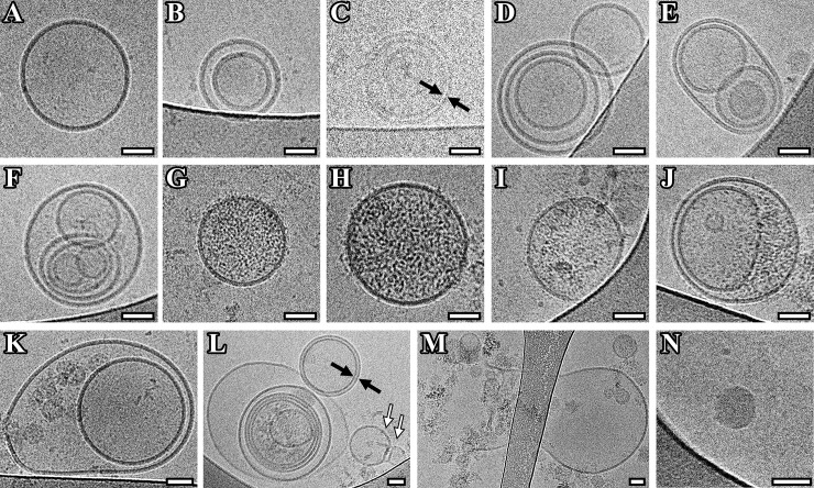
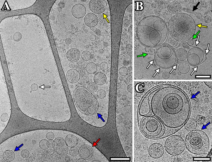
Extracellular vesicles (EVs) are membrane-enclosed vesicles which play important role for cell communication and physiology. EVs are found in many human biological fluids, including blood, breast milk, urine, cerebrospinal fluid (CSF), ejaculate, saliva etc. These nano-sized vesicles contain proteins, mRNAs, microRNAs, non-coding RNAs and lipids that are derived from producing cells. EVs deliver complex sets of biological information to recipient cells thereby modulating their behaviors by their molecular cargo. In this way EVs are involved in the pathological development and progression of many human disorders, including neurodegenerative diseases. In this study EVs purified by ultracentrifugation from CSF of patients with Parkinson's disease (PD) and individuals of the comparison group were characterized using nanoparticle tracking analysis, flow cytometry and cryo-electron microscopy. Vesicular size and the presence of exosomal marker CD9 on the surface provided evidence that most of the EVs were exosome-like vesicles. Cryo-electron microscopy allowed us to visualize a large spectrum of extracellular vesicles of various size and morphology with lipid bilayers and vesicular internal structures. Thus, we described the diversity and new characteristics of the vesicles from CSF suggesting that subpopulations of EVs with different and specific functions may exist.


Conflict of interest statement

The authors have declared that no competing interests exist.

Figures

Fig 1
Fig 1. Characterization of EVs isolated from cerebrospinal fluid.
(A) Example of Flow cytometric analysis of the CD9 exosomal marker expression on the surface of EVs isolated from CSF (EVs). Immunobeads, which were not incubated with EVs during sample preparation, were used as a negative control ((–) control). The exosomal standard included in the HansaBioMed exosome cytometric assay kit was used as a positive control ((+) control); (B) Nanoparticle tracking analysis (NTA) of particle size and concentration in the sample of vesicles isolated from pooled CSF of PD patients.
Fig 2
Fig 2. Representation in size of EVs with various morphology.
Single, double, double membrane, multilayer and vesicles with electron dense cargo were visualized by cryo-EM both in vesicle samples of pooled CSF from (A) PD patients and (B) individuals of comparison group. N is the number of particles of the indicated morphological type.
Fig 3
Fig 3. Cryo-EM images of EVs isolated from pooled CSF of Parkinson's disease patients.
(A) Single vesicles; (B, K) double vesicles; (C, L) double-membrane vesicles; (C-F, L) multilayer vesicles; (G-J, L) vesicles with electron dense cargo in lumen; (K, M) vesicles with broken membrane. The arrows point to vesicle with double membrane (black arrow) and “bowling pin” vesicle (white arrow). Scale bars are 50 nm.
Fig 4
Fig 4. Cryo-EM images of EVs isolated from pooled CSF sample from individuals of the comparison group.
(A) Micrograph of single (white arrow), double (red arrow), multilayer (blue) vesicles and vesicles with electron dense cargo in lumen (yellow arrow); (B) micrograph of double-membrane vesicles (green arrow), vesicles with electron dense cargo in lumen (yellow arrow), vesicle in form of sack (white arrows), distinctly elongated vesicle (black arrow); (C) micrograph of multilayer vesicles (blue arrows). Scale bars are 200 nm for figures A, B and 100 nm for figure C.

References

    1. Raposo G, Stoorvogel W. Extracellular vesicles: exosomes, microvesicles, and friends. J Cell Biol. 2013; 200(4): 373–83. 10.1083/jcb.201211138 - DOI - PMC - PubMed
    1. Tofaris GK. A Critical Assessment of Exosomes in the Pathogenesis and Stratification of Parkinson's Disease. J Parkinsons Dis. 2017; 7(4): 569–576. 10.3233/JPD-171176 - DOI - PMC - PubMed
    1. Greening DW, Simpson RJ. Understanding extracellular vesicle diversity—current status. Expert Rev Proteomics. 2018; 15(11): 887–910. 10.1080/14789450.2018.1537788 . - DOI - PubMed
    1. Clemmens H, Lambert DW. Extracellular vesicles: translational challenges and opportunities. Biochem Soc Trans. 2018; 46(5): 1073–1082. 10.1042/BST20180112 . - DOI - PubMed
    1. Isola AL, Chen S. Exosomes: The Messengers of Health and Disease. Curr Neuropharmacol. 2017; 15(1): 157–165. 10.2174/1570159X14666160825160421 - DOI - PMC - PubMed

Publication types

MeSH terms