

Fertility-preserving local excision under a hysteroscope with combined chemotherapy in a 6-year-old child with clear cell adenocarcinoma of the cervix: A case report and review of the literature
- PMID: 32000369
- PMCID: PMC7004716
- DOI: 10.1097/MD.0000000000018646
Fertility-preserving local excision under a hysteroscope with combined chemotherapy in a 6-year-old child with clear cell adenocarcinoma of the cervix: A case report and review of the literature
Abstract
Introduction: Clear cell adenocarcinoma of the cervix (CCAC), a rare and more severe type of gynecological cancer, is especially rare in pediatric patients. Traditionally, surgery following chemotherapy (CT) and radiation therapy is the preferred treatment for CCAC; however, patients have poor 5-year survival rates than other types of cervical cancers.
Patient concerns: A 6-year-old girl with a history of vaginal discharge for 18 months was diagnosed with CCAC by histological examination. Her parents refused the traditional treatment of radical hysterectomy and lymph node dissection because of her young age.
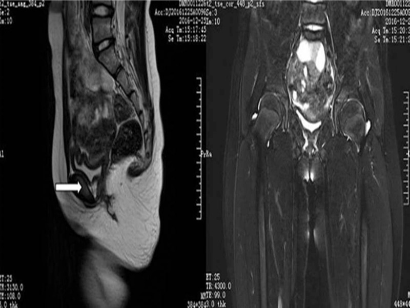
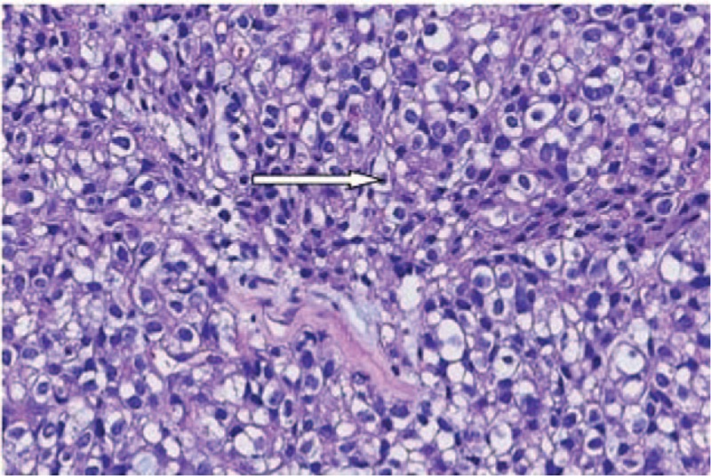
Diagnosis: The patient's tests revealed negative human papilloma virus and negative methylated paired box 1 gene results. The tumor mass histopathology revealed stage IIA1 CCAC that originated from the cervix.
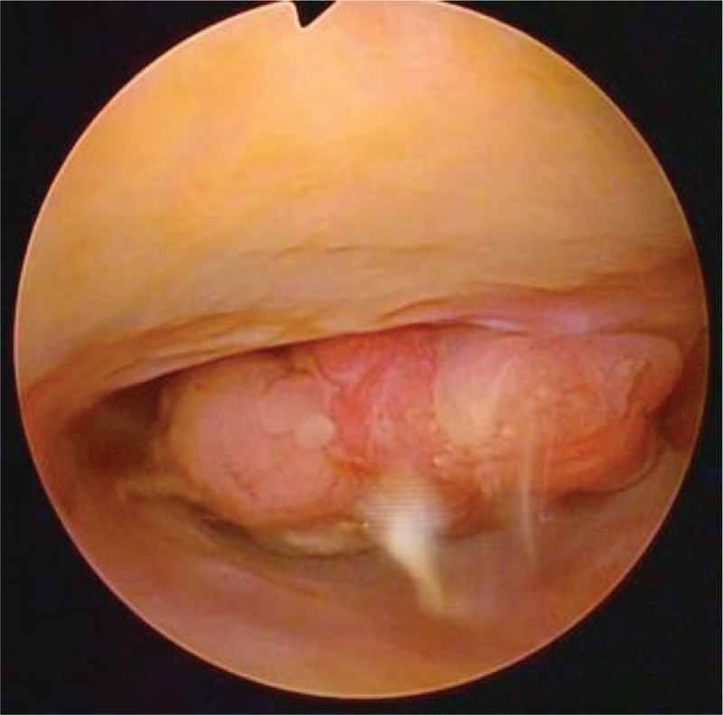
Interventions: Tumor mass excision with preservation of the cervix by electrosurgical biopsy under hysteroscopy was performed. Four cycles of docetaxel and oxaliplatin CT were administered every 3 weeks.
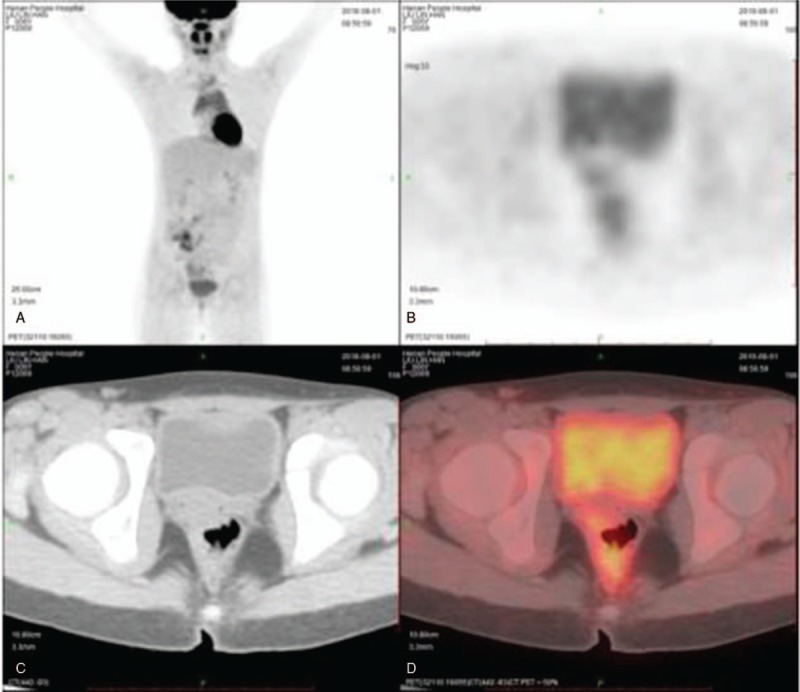
Outcomes: No signs of recurrence were observed in the 28 months after final treatment and diagnosis on magnetic resonance imaging, color ultrasonic imaging, and gynecological examination. Serologic tumor biomarkers were also within normal ranges.
Conclusions: This is the first reported CCAC case in which the primary treatment included electrosurgical biopsy of the polypoid mass under hysteroscopy, followed by CT without traditional treatment: radical surgery with pelvic and/or lymphadenectomy for fertility preservation. This is a new treatment approach for young CCAC patients without the use of surgery.
Conflict of interest statement
The authors have no conflicts of interest to disclose.
Figures
Similar articles
-
A young woman with clear cell adenocarcinoma of the uterine cervix.Int J Clin Oncol. 2003 Dec;8(6):399-404. doi: 10.1007/s10147-003-0358-0. Int J Clin Oncol. 2003. PMID: 14663645
-
Cervical clear cell carcinoma: Case report and literature review.Medicine (Baltimore). 2024 Mar 29;103(13):e37449. doi: 10.1097/MD.0000000000037449. Medicine (Baltimore). 2024. PMID: 38552088 Free PMC article. Review.
-
Oncological and pregnancy outcomes after high-dose density neoadjuvant chemotherapy and fertility-sparing surgery in cervical cancer.Gynecol Oncol. 2014 Nov;135(2):213-6. doi: 10.1016/j.ygyno.2014.08.021. Epub 2014 Aug 23. Gynecol Oncol. 2014. PMID: 25159484
-
[Clear Cell Carcinoma of Cervix in a 12-Year-Old Girl: A Case Report].Sichuan Da Xue Xue Bao Yi Xue Ban. 2021 May;52(3):534-538. doi: 10.12182/20210560208. Sichuan Da Xue Xue Bao Yi Xue Ban. 2021. PMID: 34018378 Free PMC article. Chinese.
-
Mullerian adenosarcoma of the cervix in a 10-year-old girl: case report and review of the literature.J Pediatr Adolesc Gynecol. 2009 Aug;22(4):e45-51. doi: 10.1016/j.jpag.2008.06.001. Epub 2009 Jun 2. J Pediatr Adolesc Gynecol. 2009. PMID: 19493521 Review.
Cited by
-
Primary Clear Cell Adenocarcinoma of the Uterine Cervix in a 14-Year-Old Virgin Girl: Case Report.Int J Environ Res Public Health. 2022 Dec 11;19(24):16652. doi: 10.3390/ijerph192416652. Int J Environ Res Public Health. 2022. PMID: 36554533 Free PMC article.
-
Significance of Magnetic Resonance Imaging Combining with Detection of Serum HE4, TSGF, and CD105 Levels in Diagnosis and Treatment of Moderate to Advanced Cervical Cancer.Contrast Media Mol Imaging. 2022 Feb 24;2022:2090654. doi: 10.1155/2022/2090654. eCollection 2022. Contrast Media Mol Imaging. 2022. PMID: 39281827 Free PMC article.
-
Fertility-sparing radical resection of juvenile clear cell adenocarcinoma of the cervix by pneumovaginal endoscopic surgery.Gynecol Oncol Rep. 2023 Jan 18;45:101135. doi: 10.1016/j.gore.2023.101135. eCollection 2023 Feb. Gynecol Oncol Rep. 2023. PMID: 36714371 Free PMC article.
-
Factors Associated with Patient Survival in Clear Cell Adenocarcinoma of the Cervix: A Single-Center Experience in China.Int J Gen Med. 2022 May 3;15:4625-4634. doi: 10.2147/IJGM.S358094. eCollection 2022. Int J Gen Med. 2022. PMID: 35535144 Free PMC article.
-
Clear Cell Carcinoma of the Uterine Cervix in a 14-Year-Old Girl: A Case Report.Cureus. 2025 Jan 7;17(1):e77060. doi: 10.7759/cureus.77060. eCollection 2025 Jan. Cureus. 2025. PMID: 39917107 Free PMC article.
References
-
- Bray F, Ferlay J, Soerjomataram I, et al. Global cancer statistics 2018: GLOBOCAN estimates of incidence and mortality worldwide for 36 cancers in 185 countries. CA Cancer J Clin 2018;68:394–424. - PubMed
-
- Choi SJ, Kim JE, Kim HS, et al. Clear cell adenocarcinoma of the uterine cervix in a 15-year-old girl: a case report. Korean J Radiol 2013;69:321–5.
-
- Premarket Assessment of Pediatric Medical Devices: Guidance for Industry and FDA Staff. U.S. Food and Drug Administration. Available at: https://www.fda.gov/downloads/MedicalDevices/DeviceRegulationandGuidance.... Published March 24, 2014. Updated September 10, 2018. Accessed.
-
- Reich O, Tamussino K, Lahousen M, et al. Clear cell carcinoma of the uterine cervix: pathology and prognosis in surgically treated stage IB-IIB disease in women not exposed in utero to diethylstilbestrol. Gynecol Oncol 2000;76:331–5. - PubMed
