

Preoperative assessment of contrast-enhanced spectral mammography of diagnosed breast cancers after sonographic biopsy: Correlation to contrast-enhanced magnetic resonance imaging and 5-year postoperative follow-up
- PMID: 32000448
- PMCID: PMC7004697
- DOI: 10.1097/MD.0000000000019024
Preoperative assessment of contrast-enhanced spectral mammography of diagnosed breast cancers after sonographic biopsy: Correlation to contrast-enhanced magnetic resonance imaging and 5-year postoperative follow-up
Abstract
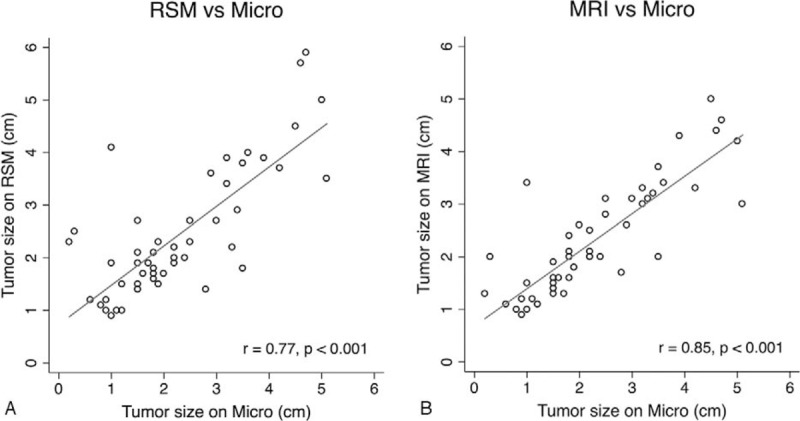
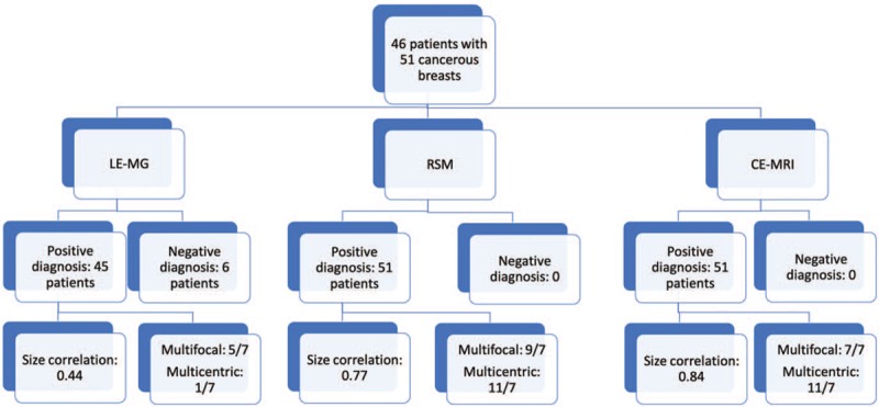
To assess the feasibility of using contrast-enhanced spectral mammography (CESM) for operative planning of patients with breast cancers who were initially diagnosed by sonographic guided biopsy.With the approval of the Institutional Review Board of our hospital, we retrospectively reviewed the data on patients with breast cancers who underwent CESM and contrast-enhanced magnetic resonance imaging (CE-MRI) prior to operation and were followed up for at least 5 years postoperatively. The patients with breast cancer diagnosed by sonographic guided biopsy without mammography were included for analysis. The size and number of cancers on low-energy mammograms (LE-MG), recombined subtracted mammograms (RSM), and CE-MRI were recorded and compared with microscopic histopathologic data and at least 5 years of clinical follow-up data.Fifty-one cancerous breasts of 46 patients were included in the analysis. All the principal cancers could be detected by RSM or CE-MRI; however, only 45 were by LE-MG. The Pearson correlation coefficients for the size on microscopy were 0.44 for LE-MG, 0.77 for RSM, and 0.84 for CE-MRI (all P-values ≤.001). Regarding the microscopic reports, RSM or CE-MRI had sensitivities of 100% and a positive predictive value of 63.6% for multicentric cancers. One breast cancer with partial mastectomy recurred after 3 years of follow-up.CESM was feasible for assessing the cancer extension and multicentric cancers as secondary examination in patients with diagnosed breast cancers after sonographic biopsy.
Conflict of interest statement
The authors have no conflicts of interest to disclose.
Figures
References
-
- Jatoi I, Proschan MA. Randomized trials of breast-conserving therapy versus mastectomy for primary breast cancer: a pooled analysis of updated results. Am J Clin Oncol 2005;28:289–94. - PubMed
-
- Anastassiades O, Iakovou E, Stavridou N, et al. Multicentricity in breast cancer. A study of 366 cases. Am J Clin Pathol 1993;99:238–43. - PubMed
-
- Pisano ED, Gatsonis C, Hendrick E, et al. Diagnostic performance of digital versus film mammography for breast-cancer screening. N Engl J Med 2005;353:1773–83. - PubMed
-
- Kolb TM, Lichy J, Newhouse JH. Comparison of the performance of screening mammography, physical examination, and breast US and evaluation of factors that influence them: an analysis of 27,825 patient evaluations. Radiology 2002;225:165–75. - PubMed
MeSH terms
Substances
LinkOut - more resources
Full Text Sources
Medical
