

To treat or not to treat: a clinical series of retinal arterial macroaneurysms: A single-center retrospective study
- PMID: 32000459
- PMCID: PMC7004597
- DOI: 10.1097/MD.0000000000019077
To treat or not to treat: a clinical series of retinal arterial macroaneurysms: A single-center retrospective study
Abstract
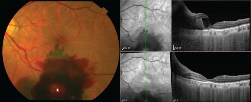
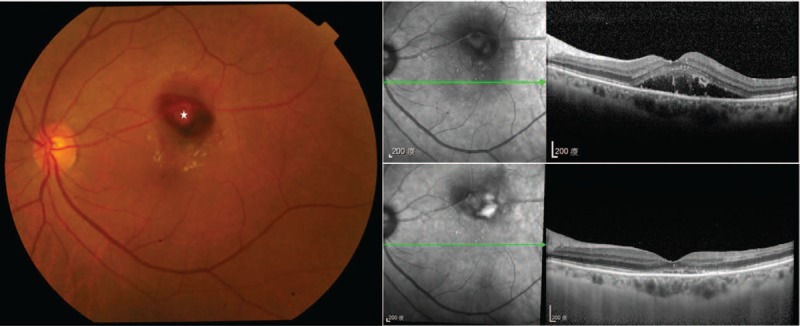
Retinal arterial macroaneurysms (RAMs) develop as outpouchings of the arterial wall that is weakened by arteriosclerosis. The traditional treatment of RAMs comprises observation, focal laser photocoagulation, or surgery. Recently, intravitreal injection of anti-vascular endothelial growth factor (VEGF) drugs has been announced as an effective therapy for fovea-threatening RAMs and quickly improve visual acuity and central retinal thickness (CRT).In the retrospective series, medical charts and ocular images of 24 patients diagnosed as having RAM between May 2011 and November 2018 in our facility were reviewed to delineate clinical manifestations and visual prognosis in RAM patients receiving different treatment modalities. Twenty-four patients (25 eyes; 11 men and 13 women) were enrolled, and one eye with comorbidity of branch retinal vein occlusion was excluded. The mean age of the patients was 69.00 ± 13.45 years. Fourteen patients (58.33%) had a history of hypertension, and 17 patients (70.83%) were aged > 60 years. Furthermore, patients with fovea-threatening RAMs presented with either hypertension or were aged > 60 years.Eyes with fovea involvement (n = 18) were analyzed and separated into two groups according to their treatment modalities: those receiving anti-VEGF intravitreal injections (n = 13) and observation only (n = 5). The baseline visual acuity revealed no significant difference in the two groups. In patients receiving anti-VEGF intravitreal injections, a significantly better visual acuity was detected after anti-VEGF intravitreal injections than the baseline visual acuity (logMAR, 0.78 ± 0.51 vs 1.52 ± 0.48, P < .001), and CRT significantly improved (505.50 ± 159.26 μm vs 243.60 ± 60.17 μm, P = .001). Patients receiving anti-VEGF intravitreal injections also revealed better final visual acuity than those in the observation group (logMAR, 0.78 ± 0.51 vs 1.34 ± 0.48, P = .04).A systematic work-up for hypertension and arteriosclerotic disease could be considered the recommended procedure once RAM has been diagnosed. With better final visual acuity, significant visual improvements, and fast reduction of CRT observed in patients with fovea-threatening RAMs receiving anti-VEGF intravitreal injections, intravitreal anti-VEGF was considered an effective therapy for complicated RAM. During the follow-up period, the majority of RAM eyes had good maintenance of visual function even with foveal complications.
Figures
References
-
- Pitkanen L, Tommila P, Kaarniranta K, et al. Retinal arterial macroaneurysms. Acta Ophthalmol 2014;92:101–4. - PubMed
-
- Lee EK, Woo SJ, Ahn J, et al. Morphologic characteristics of retinal arterial macroaneurysm and its regression pattern on spectral-domain optical coherence tomography. Retina (Philadelphia, PA) 2011;31:2095–101. - PubMed
-
- Moosavi RA, Fong KC, Chopdar A. Retinal artery macroaneurysms: clinical and fluorescein angiographic features in 34 patients. Eye (London, England) 2006;20:1011–20. - PubMed
MeSH terms
Substances
LinkOut - more resources
Full Text Sources
Other Literature Sources
Research Materials
