

Utility of Contrast-Enhanced Harmonic Endoscopic Ultrasound for the Guidance and Monitoring of Endoscopic Radiofrequency Ablation
- PMID: 32000466
- PMCID: PMC7667920
- DOI: 10.5009/gnl19123
Utility of Contrast-Enhanced Harmonic Endoscopic Ultrasound for the Guidance and Monitoring of Endoscopic Radiofrequency Ablation
Abstract
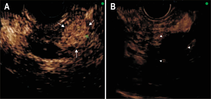
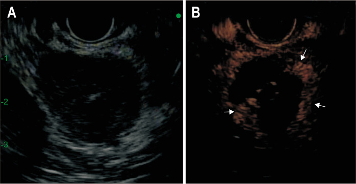
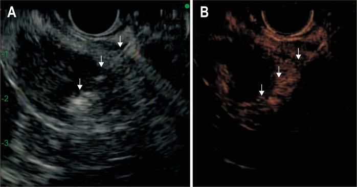
Background/aims: Interventional endoscopists may utilize contrast-enhanced harmonic endoscopic ultrasound (CEHEUS) for image guidance during radiofrequency ablation (RFA) because of its capability to delineate real-time tumor perfusion dynamics. The purpose of this study was to assess the utility of CEH-EUS for the guidance and monitoring of endoscopic RFA.
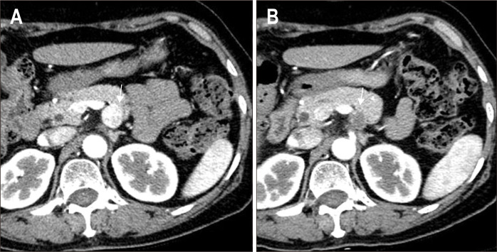
Methods: Nineteen consecutive patients with solid abdominal tumors who underwent CEH-EUS and endoscopic RFA were included. The extent of the ablation was assessed by CEH-EUS at 5 to 7 days after RFA. Additional RFAs were performed under CEH-EUS guidance.
Results: The diagnoses were as follows: nonfunctioning neuroendocrine tumor (n=13), solid pseudopapillary neoplasm (SPN) (n=2), insulinoma (n=1), left adrenal adenoma (n=2), and left adrenal oligometastasis (n=1). Pre-CEH-EUS findings revealed that 17 cases showed hyperenhanced patterns and two cases of SPN showed isoenhanced patterns. CEH-EUS-assisted RFA was technically feasible in all 19 patients. After the first RFA session, seven patients of the treated tumors showed the disappearance of intratumoral enhancement on CEH-EUS, whereas 12 showed residual contrast enhancement. Twelve patients with incomplete ablation were further treated with additional RFA under real-time CEH-EUS guidance. Radiologic complete response was observed in 13 patients (68.4%). Among the 35 ablation procedures, the only adverse events were two episodes of pancreatitis (5.7%; 1 moderate and 1 mild). During the median follow-up of 28 months, the local recurrence rate was 7.7%.
Conclusions: The application of CEH-EUS for RFA could be helpful in assessing early treatment response after ablation and targeting residual viable tumors during additional ablation sessions.
Keywords: Contrast agent; Endosonography; Radiofrequency ablation.
Conflict of interest statement
No potential conflict of interest relevant to this article was reported.
Figures
Comment in
-
Contrast Harmonic-Enhanced Endoscopic Ultrasound (EUS) Is the Perfect Companion of EUS-Guided Tumor Ablation.Gut Liver. 2020 Sep 15;14(5):669-670. doi: 10.5009/gnl20077. Gut Liver. 2020. PMID: 32773387 Free PMC article. No abstract available.
References
MeSH terms
LinkOut - more resources
Full Text Sources
