. 2020 Jan 30;21(1):38.
doi: 10.1186/s12931-020-1299-0.

Periostin plays a critical role in the cell cycle in lung fibroblasts

Affiliations

Periostin plays a critical role in the cell cycle in lung fibroblasts

Tomohito Yoshihara et al. Respir Res. .

Abstract

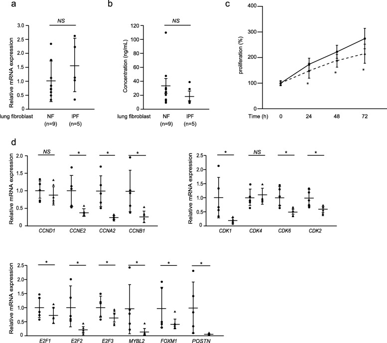
Background: Idiopathic pulmonary fibrosis (IPF) is a devastating disease with a median survival of only three to 5 years. Fibroblast proliferation is a hallmark of IPF as is secretion of extracellular matrix proteins from fibroblasts. However, it is still uncertain how IPF fibroblasts acquire the ability to progressively proliferate. Periostin is a matricellular protein highly expressed in the lung tissues of IPF patients, playing a critical role in the pathogenesis of pulmonary fibrosis. However, it remains undetermined whether periostin affects lung fibroblast proliferation.

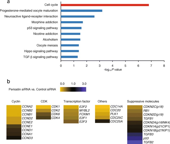
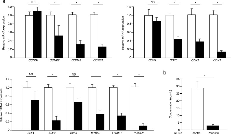
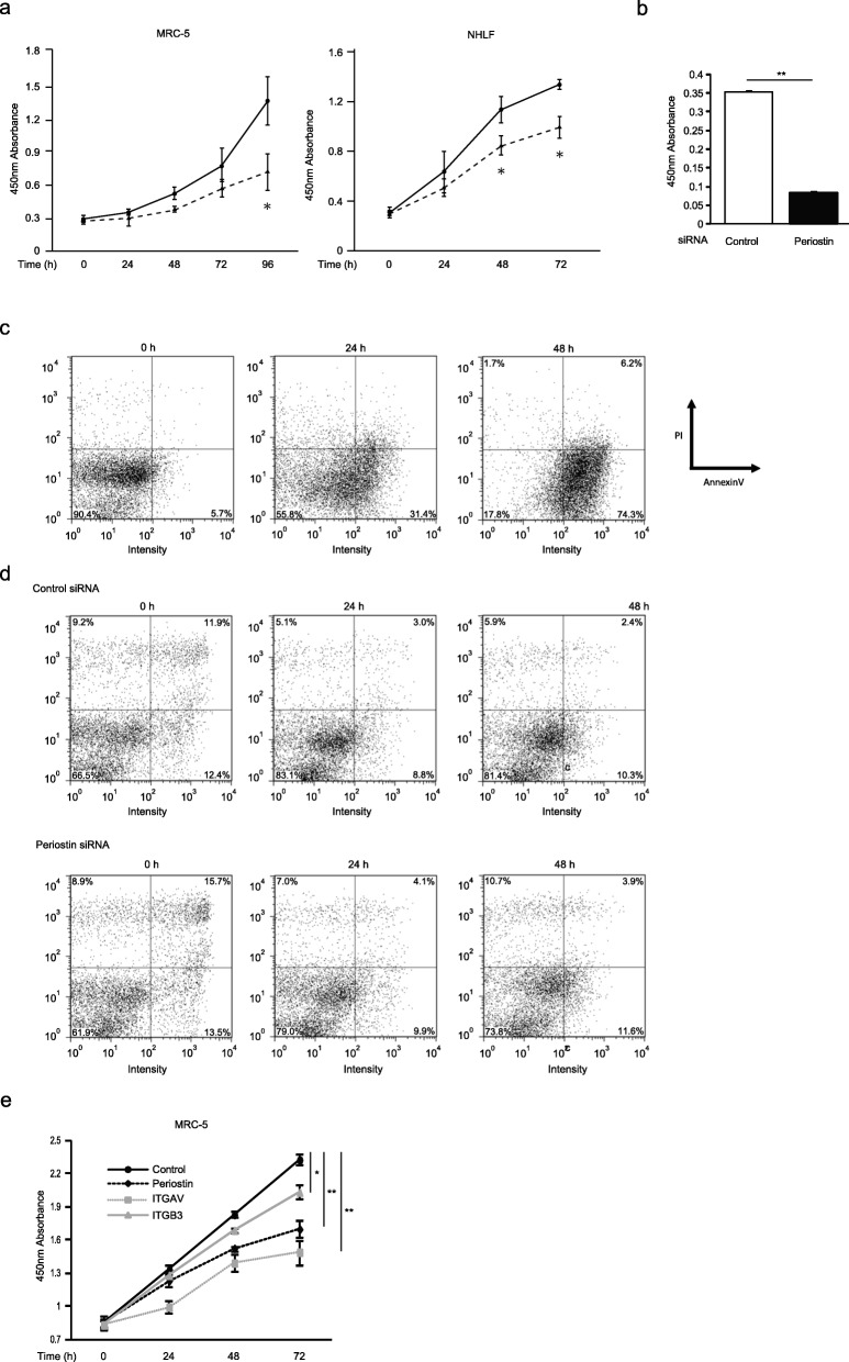
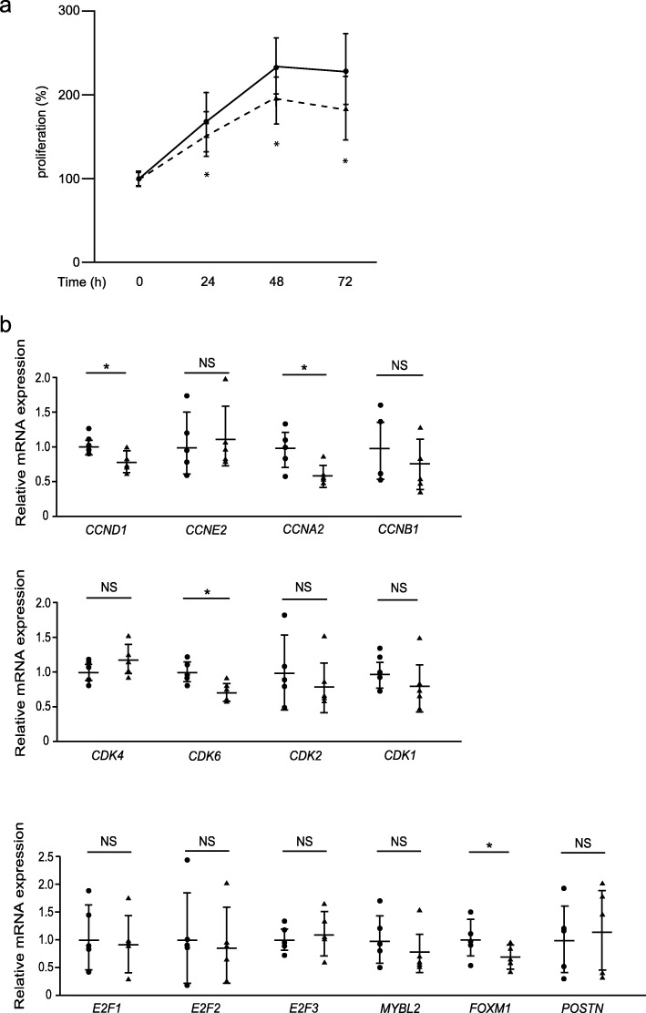
Methods: In this study, we first aimed at identifying periostin-dependently expressed genes in lung fibroblasts using DNA microarrays. We then examined whether expression of cyclins and CDKs controlling cell cycle progression occur in a periostin-dependent manner. We next examined whether downregulation of cell proliferation-promoting genes by knockdown of periostin or integrin, a periostin receptor, using siRNA, is reflected in the cell proliferation of lung fibroblasts. We then looked at whether lung fibroblasts derived from IPF patients also require periostin for maximum proliferation. We finally investigated whether CP4715, a potent inhibitor against integrin αVβ3 (a periostin receptor), which we have recently found blocks TGF-β signaling, followed by reduced BLM-induced pulmonary fibrosis in mice, can block proliferation of lung fibroblasts derived from IPF patients.

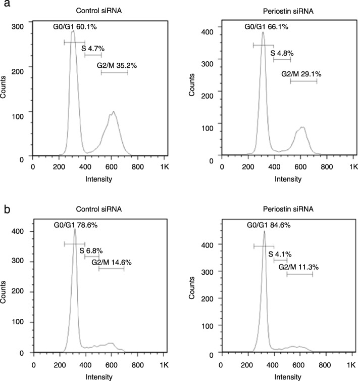
Results: Many cell-cycle-related genes are involved in the upregulated or downregulated genes by periostin knockdown. We confirmed that in lung fibroblasts, periostin silencing downregulates expression of several cell-cycle-related molecules, including the cyclin, CDK, and, E2F families, as well as transcription factors such as B-MYB and FOXM1. Periostin or integrin silencing slowed proliferation of lung fibroblasts and periostin silencing increased the distribution of the G0/G1 phase, whereas the distribution of the G2/M phase was decreased. Lung fibroblasts derived from IPF patients also required periostin for maximum proliferation. Moreover, CP4715 downregulated proliferation along with expression of cell-cycle-related genes in IPF lung fibroblasts as well as in normal lung fibroblasts.

Conclusions: Periostin plays a critical role in the proliferation of lung fibroblasts and the present results provide us a solid basis for considering inhibitors of the periostin/integrin αVβ3 interaction for the treatment of IPF patients.

Keywords: Cell cycle; Fibroblast; Idiopathic pulmonary fibrosis; Integrin; Periostin; Proliferation.


Conflict of interest statement

KA and SM are employees of Meiji Seika Pharma Co. Ltd.. The other authors declare no conflict of interest.

Figures

Fig. 1
Fig. 1
Comparison of the gene profiles affected by periostin silencing in lung fibroblasts. a MRC-5 cells with control or POSTN-specific siRNA (KD) were subjected to DNA microarray analysis. Genes downregulated by less than one third or genes upregulated by more than three-fold by knockdown of periostin were applied to DAVID analysis. Highly ranked GO terms are depicted. b Heat maps depict the genes in the GO term, cell cycle, with control siRNA versus periostin siRNA
Fig. 2
Fig. 2
Effect of periostin silencing on expression of cell cycle-related genes in lung fibroblasts. MRC-5 cells were treated with 10 nM control (open box) or periostin (closed box) siRNA. a qRT-PCR for the indicated important cell cycle progression genes were performed in periostin-silenced MRC-5 cells after 48 h. The values were adjusted by GAPDH expression, and the fold changes are shown. b Periostin protein in the supernatant after 72 h. Values are mean ± SD of three independent experiments. *P < 0.05, NS: not significant
Fig. 3
Fig. 3
Periostin silencing slows cell proliferation in lung fibroblasts. a, b, e The growth curves of MRC-5 cells or NHLF. a The cells with (dashed line) or without (solid line) treatment of periostin knockdown were plated at a density of 1.0 × 104 cells/well in 96-well plates. b The cells were treated with control siRNA or periostin siRNA for 48 h and pulsed with BrdU. After 12 h, the incorporation of BrdU was counted. Values are mean ± SD of three independent experiments. e The cells treated with siRNA for control (black solid line), periostin (black dashed line), αV integrin (gray dashed line), or β3 integrin (gray solid line) were plated at a density of 1.0 × 104 cells/well in 96-well plates. The cell numbers were evaluated at the indicated times. Values are mean ± SD of three independent experiments. *P < 0.05, **P < 0.01. c, d Flow cytometric analysis of annexin V (horizontal) and PI (vertical) labeling is depicted. The proportions of each fraction have been inserted. The same experiments were performed twice. MRC-5 cells were treated with 50 μg/mL cycloheximide and 50 ng/mL TNF-α (c) or siRNA for periostin (d) for the indicated times
Fig. 4
Fig. 4
Effect of periostin on the distribution of the cell cycle in lung fibroblasts. After MRC-5 cells and NHLFs were serum-deprived for 24 h, the cells were treated with 10 nM control or periostin siRNA for 48 h. Distribution of the cell cycle of MRC-5 cells (a) and NHLFs (b) as estimated by flow cytometry is depicted. We performed the same experiments for three times and show the representative data
Fig. 5
Fig. 5
Effect of periostin silencing on proliferation of IPF lung fibroblasts. a, b Expression of periostin RNA estimated by qRT-PCR or periostin protein estimated by the ELISA assay. The values were adjusted by GAPDH expression, and the folds for the values are shown for qRT-PCR in panel a. The complete results of nine clones of normal lung fibroblasts and five clones of IPF lung fibroblasts are depicted. Values are mean ± SD of three independent experiments. *P < 0.05. c The growth curves of IPF lung fibroblasts. The cells with (dashed line) or without (solid line) knockdown of periostin were plated at a density of 1.0 × 104 cells/well in 96-well plates. The cell numbers were evaluated at the indicated times. Values are mean ± SD of three independent experiments. *P < 0.05. d IPF lung fibroblasts were treated with 10 nM control (closed circle) or periostin (closed triangle) siRNA for 48 h. qRT-PCR for the indicated important cell cycle progression genes in periostin-silenced lung fibroblasts. The values were adjusted by GAPDH expression, and the folds for the values are shown. Values are mean ± SD of three independent experiments. *P < 0.05, NS: not significant
Fig. 6
Fig. 6
Effect of CP4715 on proliferation of IPF lung fibroblasts. a The growth curves of IPF lung fibroblasts. The cells in the absence (solid line) or presence (dashed line) of 1 μM CP4715 dissolved in DMSO were plated at a density of 1.0 × 104 cells/well in 96-well plates. Cell numbers were evaluated at the indicated times. Values are mean ± SD of three independent experiments. *P < 0.05. b IPF lung fibroblasts were treated without (closed circle) or with (closed triangle) 1 μM CP4715 dissolved in DMSO for 24 h. qRT-PCR for the indicated important cell cycle progression genes in CP4715-treated lung fibroblasts were performed. The values were adjusted by GAPDH expression, and the folds for the values are shown. Values are mean ± SD of three independent experiments. *P < 0.05, **P < 0.01, NS: not significant

References

    1. Martinez FJ, Collard HR, Pardo A, Raghu G, Richeldi L, Selman M, et al. Idiopathic pulmonary fibrosis. Nat Rev Dis Primers. 2017;3:17074. doi: 10.1038/nrdp.2017.74. - DOI - PubMed
    1. Toonkel RL, Hare JM, Matthay MA, Glassberg MK. Mesenchymal stem cells and idiopathic pulmonary fibrosis. Potential for clinical testing. Am J Respir Crit Care Med. 2013;188:133–140. doi: 10.1164/rccm.201207-1204PP. - DOI - PubMed
    1. Hsu E, Shi H, Jordan RM, Lyons-Weiler J, Pilewski JM, Feghali-Bostwick CA. Lung tissues in patients with systemic sclerosis have gene expression patterns unique to pulmonary fibrosis and pulmonary hypertension. Arthritis Rheum. 2011;63:783–794. doi: 10.1002/art.30159. - DOI - PMC - PubMed
    1. Vuga LJ, Ben-Yehudah A, Kovkarova-Naumovski E, Oriss T, Gibson KF, Feghali-Bostwick C, et al. WNT5A is a regulator of fibroblast proliferation and resistance to apoptosis. Am J Respir Cell Mol Biol. 2009;41:583–589. doi: 10.1165/rcmb.2008-0201OC. - DOI - PMC - PubMed
    1. Lee JU, Cheong HS, Shim EY, Bae DJ, Chang HS, Uh ST, et al. Gene profile of fibroblasts identify relation of CCL8 with idiopathic pulmonary fibrosis. Respir Res. 2017;18:3. doi: 10.1186/s12931-016-0493-6. - DOI - PMC - PubMed

MeSH terms

Substances