Review
. 2020 Jan 30;18(1):23.
doi: 10.1186/s12951-019-0556-1.

Biodegradable polymer nanocomposites for ligament/tendon tissue engineering

Affiliations
Review

Biodegradable polymer nanocomposites for ligament/tendon tissue engineering

Magda Silva et al. J Nanobiotechnology. .

Abstract

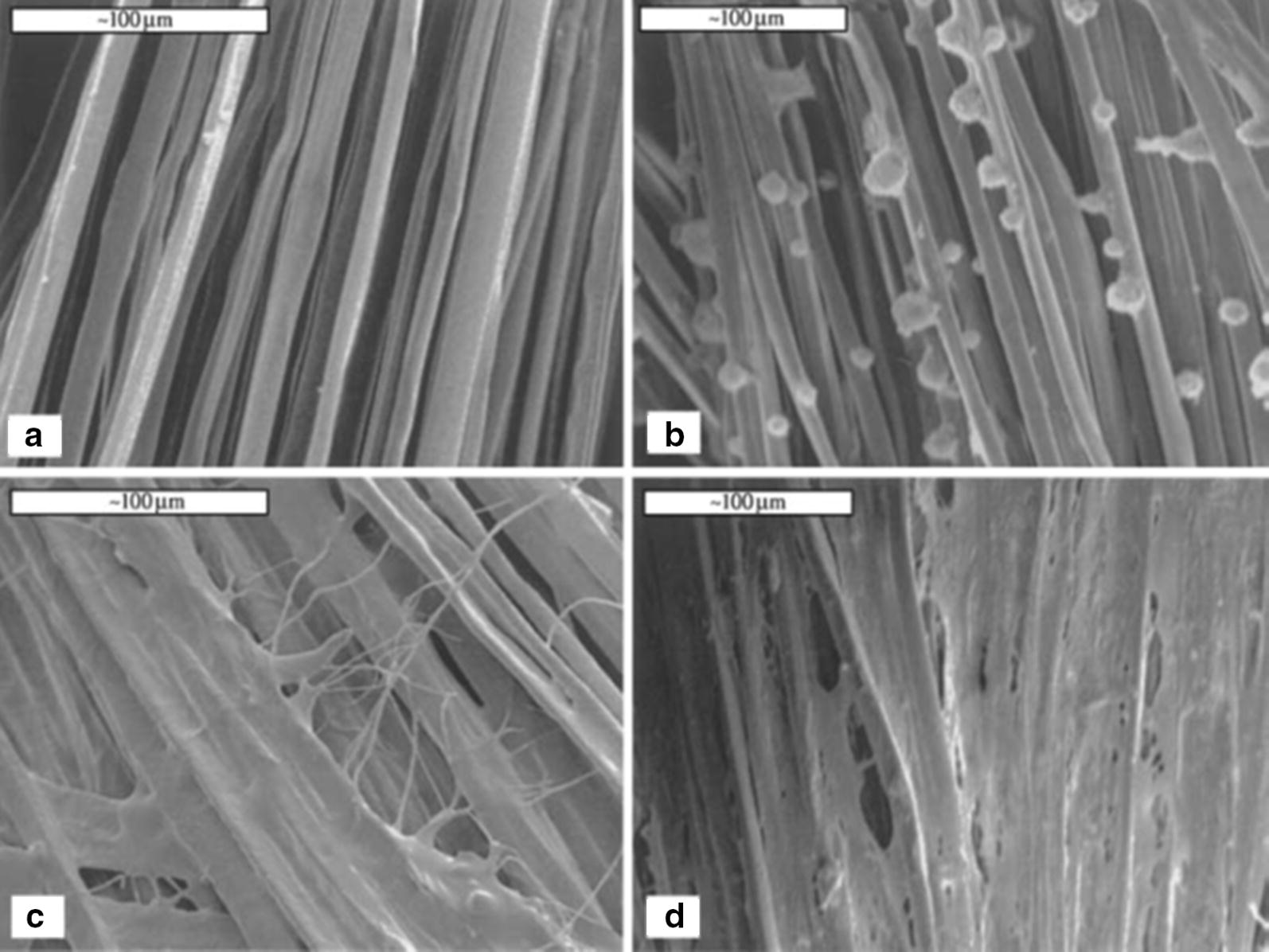
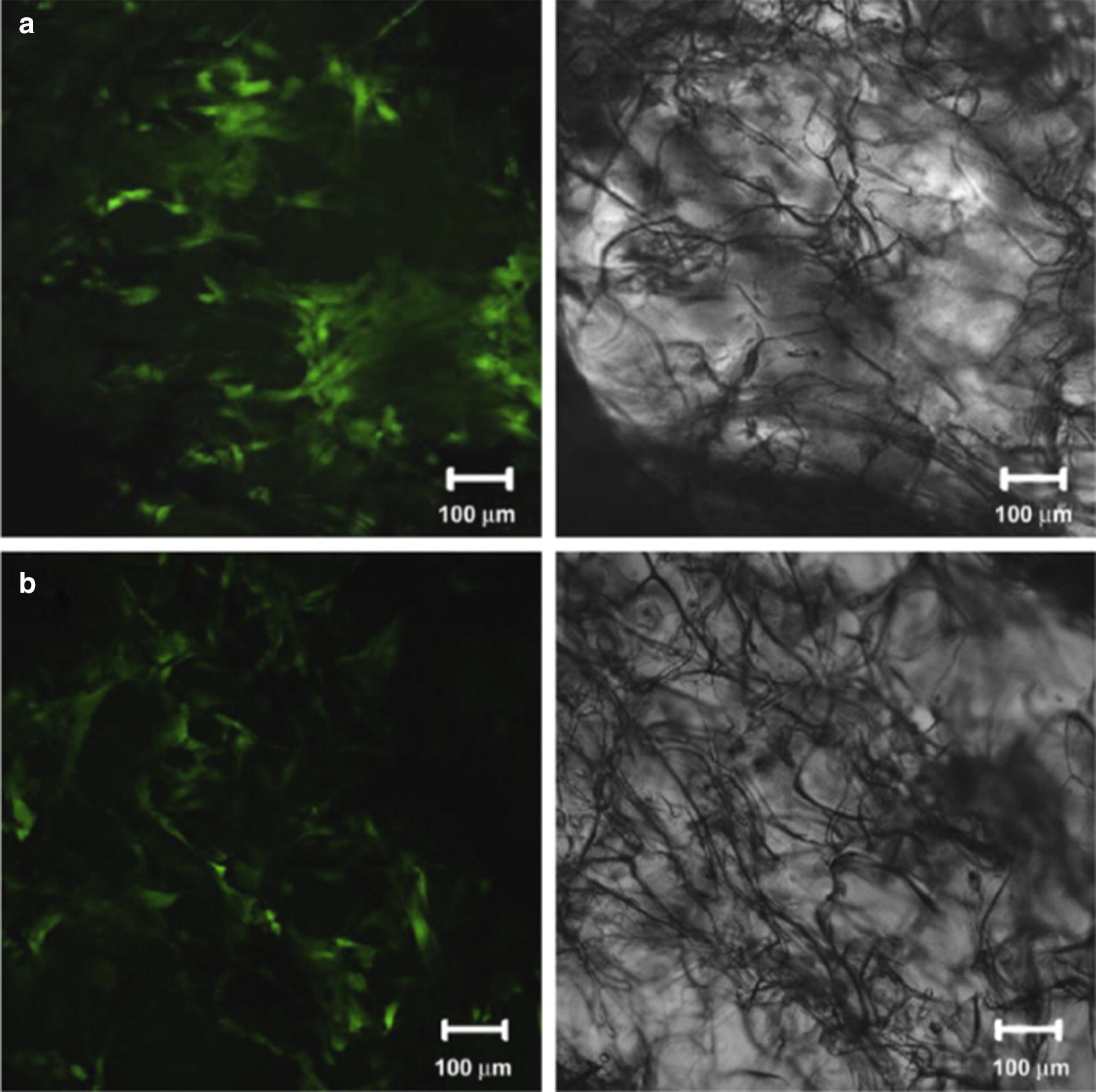
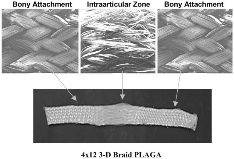
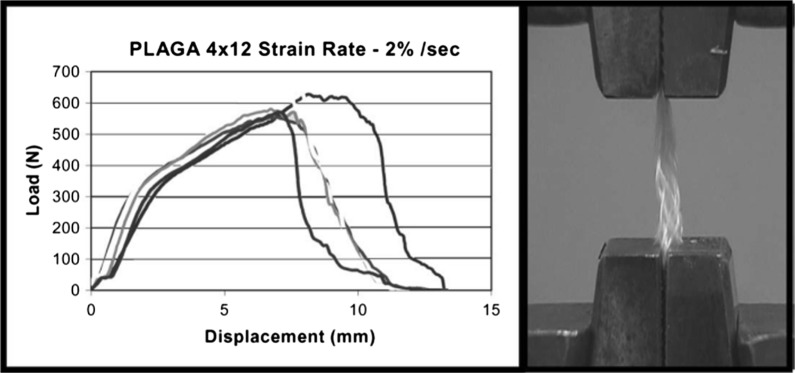
Ligaments and tendons are fibrous tissues with poor vascularity and limited regeneration capacity. Currently, a ligament/tendon injury often require a surgical procedure using auto- or allografts that present some limitations. These inadequacies combined with the significant economic and health impact have prompted the development of tissue engineering approaches. Several natural and synthetic biodegradable polymers as well as composites, blends and hybrids based on such materials have been used to produce tendon and ligament scaffolds. Given the complex structure of native tissues, the production of fiber-based scaffolds has been the preferred option for tendon/ligament tissue engineering. Electrospinning and several textile methods such as twisting, braiding and knitting have been used to produce these scaffolds. This review focuses on the developments achieved in the preparation of tendon/ligament scaffolds based on different biodegradable polymers. Several examples are overviewed and their processing methodologies, as well as their biological and mechanical performances, are discussed.

Keywords: Biodegradability; Nanocomposites; Tendon/ligament tissue engineering.


Conflict of interest statement

The authors declare that they have no competing interests.

Figures

Fig. 1
Fig. 1
a and b Structure of a twisted or cable yarn. Fibers are combined to form bundles, bundles to form strands, and strands to form cords. Yarns were labeled: A(a) × B(b) × C(c), where A, B, C represent the number of fibers/bundles/strands in the final structure, respectively and a, b, c is the number of turns per inch on each of the hierarchical levels (Adapted from [52])
Fig. 2
Fig. 2
Scanning electron microscopy (SEM) showing adherence, proliferation and cell sheet formation by human BMSCs on the silk cord matrix prior to seeding (a), time 0 following seeding (b), 1 day (c), and 14 days (d). Scale bars = 100 mm (Reprinted with permission from [16])
Fig. 3
Fig. 3
Fluorescence images of implants with silk scaffolds-with BMSCs (a) and ACL fibroblasts (b) at 4 weeks post implantation. Scale bars = 100 mm (Reprinted with permission from [56])
Fig. 4
Fig. 4
General configuration of ligament scaffold design for 3-D rectangular braid, with 3 regions: femoral tunnel attachment site, ligament region, and tibial tunnel attachment site (Reprinted with permission from [6])
Fig. 5
Fig. 5
Load-deformation curve and photomicrograph of mechanical failure of the 4 × 12 PLGA 3-D rectangular braids at a strain rate of 2%/s (Reprinted with permission from [6])
Fig. 6
Fig. 6
The cellular proliferation after culturing for 3, 7, 14 and 21 days on 5 × 5 PLLA 3D square braided scaffolds. The temporal cell growth of the ligament cells was slower as compared to the tendon cells [74]
Fig. 7
Fig. 7
ACL fibroblast on braided scaffolds after 14 days of culture. Cells grown on braided scaffolds pre-coated with Fn elaborates a great amount of matrix compared to PLGA or PLLA scaffolds without Fn. Degradation of the PGA scaffold after 2 weeks of culture resulted in extensive cell loss and matrix depletion (Reprinted with permission [72])
Fig. 8
Fig. 8
Statistical evaluation of differences in failure load (a) and stiffness (b) between the autograft group and scaffold group at 4 and 16 weeks postoperatively. *Significant difference between groups [102]
Fig. 9
Fig. 9
Commercially available cell counting kit-8 (CCK-8) result of MSCs cultured on the random nanofibrous scaffolds, aligned nanofibrous and NRSs for up to 28 days. The data are expressed as the mean ± SD. The samples marked with (*) has a significant difference between the two groups (P < 0.05) (Reprinted with permission from [106])
Fig. 10
Fig. 10
Average parameters obtained from tensile testing to failure of each region (n = 9) and the whole scaffold (n = 10). a Young’s modulus, b Ultimate tensile strength, c Strain at failure. +, #, @ indicate statistical significance with P < 0.05 (Reprinted with permission from [108])
Fig. 11
Fig. 11
Mass loss of knitted structure during 20 weeks [35]
Fig. 12
Fig. 12
Viability (a) and proliferation (b) of fibroblasts seeded in different composites after 24 and 72 h in culture. Results are normalized with respect to the values for cells cultured in PLA control (Reprinted with permission from [111])
Fig. 13
Fig. 13
Preparation of a composite tendon scaffold. The scaffold was composed of an inner part of PGA unwoven fibers (a) and an outer part of a net knitted with PGA/PLA fibers in a ratio of 4:2 (b). The outcome of assembled two parts (c) (Reprinted with permission from [115])
Fig. 14
Fig. 14
Quantification of collagen fibril diameter of in vivo engineered tendons with native tendon as a control. Collagen fibril diameter of in vivo engineered tendons increased with time. There was significant difference between 12 and 21 weeks, between 21 and 45 weeks and between 12 and 45 weeks of the AMSCs seeded group (*P < 0.001). There was significant difference between two groups at 45 weeks post implantation (*P < 0.001). Exp experimental group, Ctrl control group, w week, NRAT normal rabbit tendon (Reprinted with permission from [115])
Fig. 15
Fig. 15
SEM images of random nanofiber meshes and aligned nanofiber bundles of (a, b) PCL/CHI and (c, d) PCL/CHI/ cellulose nanocrystals (3wt.%) with the respective 2D-fast Fourier transform frequency plots. Scale bar 1 μm (Reprinted with permission from [83])
Fig. 16
Fig. 16
BMSCs-seeded (7 days of culture) scaffold produced by electrospinning bFGF-PLGA fibers onto the surfaces of knitted microfibrous silk scaffolds [105]
Fig. 17
Fig. 17
Scaffolds for ligament tissue engineering. a Braided scaffold with a fibrous intra-articular zone terminated at each end by a less porous bony attachment zones in a single braid; b twisted fibrous scaffold; c silk scaffold produced by rolling up the porous knitted silk mesh around a silk cord (Reprinted with permission from [34])
Fig. 18
Fig. 18
a Morphology of PLCL scaffold and PLCL scaffold modified with poly-l-lysine and HA by scanning electron microscopy (SB: scaffold blank; SP: PLCL-poly-l-lysine; S1L: PLCL-poly-l-lysine/HA-PLCL-poly-l-lysine). b Global structure of the multi-layer braided scaffold. The six different constitutive layers, made of 16 fibers/layer, are represented with different colors (Reprinted with permission from [69])
Fig. 19
Fig. 19
Optical microscopy picture of the ligament tissue engineering scaffold (Reprinted with permission from The Royal Society of Chemistry [123])
Fig. 20
Fig. 20
a 3D view of the theoretical designed PLA screw-like scaffold structure. b The prepared PLA screw-like scaffold. c The SEM image of the PLA scaffold surface with well-defined orthogonal structure (Reprinted with permission from [114])

References

    1. Yilgor C, Huri P, Huri G. Tissue engineering strategies in ligament regeneration. Stem Cells Int. 2012;2012:374676. doi: 10.1155/2012/374676. - DOI - PMC - PubMed
    1. Rodrigues MT, Reis RL, Gomes ME. Engineering tendon and ligament tissues: present developments towards successful clinical products. J Tissue Eng Regen Med. 2013;7:673–686. doi: 10.1002/term.1459. - DOI - PubMed
    1. Kuo CK, Marturano JE, Tuan RS. Novel strategies in tendon and ligament tissue engineering: advanced biomaterials and regeneration motifs. BMC Sports Sci Med Technol. 2010;2:20. - PMC - PubMed
    1. Tovar N, Bourke S, Jaffe M, Murthy NS, Kohn J, Gatt C, et al. A comparison of degradable synthetic polymer fibers for anterior cruciate ligament reconstruction. J Biomed Mater Res A. 2010;93:738–747. - PMC - PubMed
    1. Nau T, Teuschl A. Regeneration of the anterior cruciate ligament: current strategies in tissue engineering. World J Orthop. 2015;6:127–136. doi: 10.5312/wjo.v6.i1.127. - DOI - PMC - PubMed

LinkOut - more resources