Review
. 2020 Jan 30;19(1):19.
doi: 10.1186/s12943-020-1144-6.

Predictive biomarkers and mechanisms underlying resistance to PD1/PD-L1 blockade cancer immunotherapy

Affiliations
Review

Predictive biomarkers and mechanisms underlying resistance to PD1/PD-L1 blockade cancer immunotherapy

Daixi Ren et al. Mol Cancer. .

Erratum in

Abstract

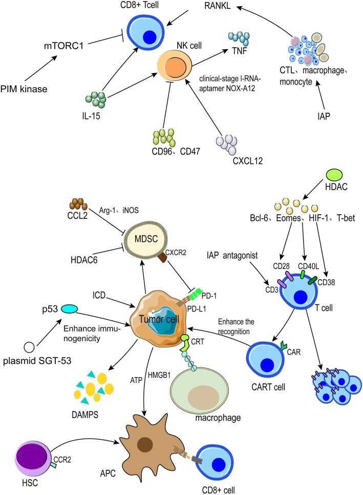
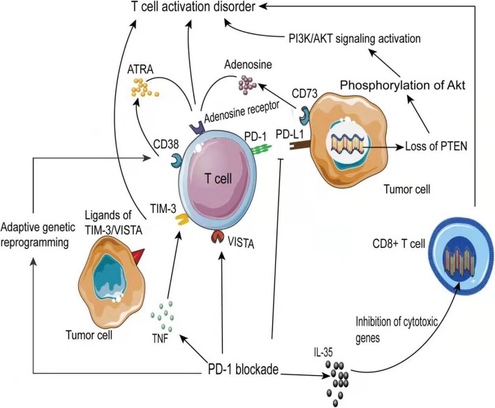
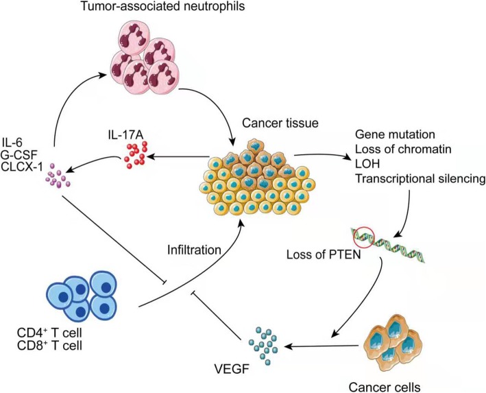
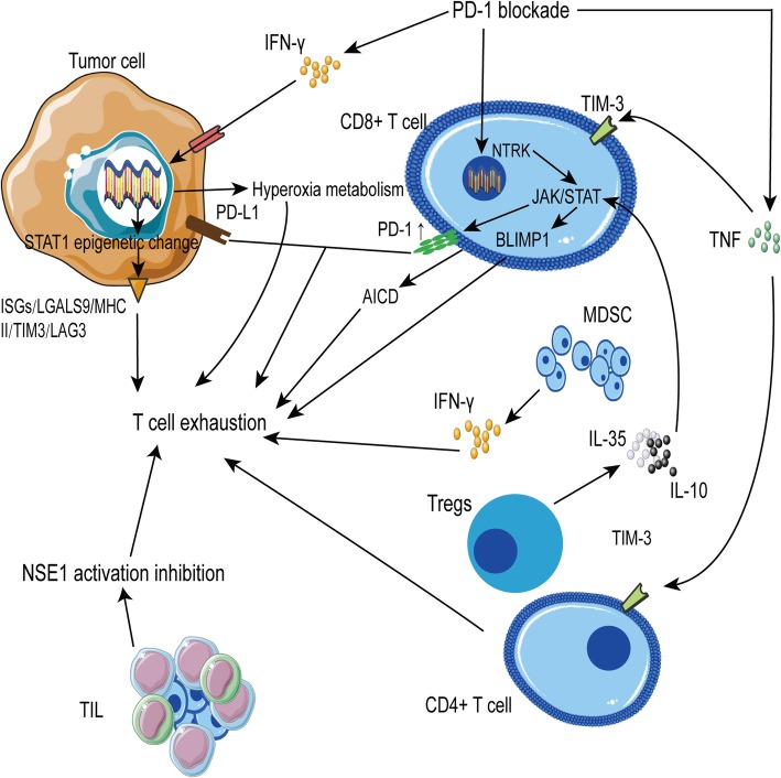
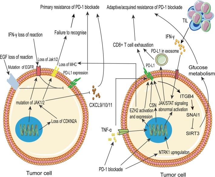
Immune checkpoint blockade targeting PD-1/PD-L1 has promising therapeutic efficacy in a variety of tumors, but resistance during treatment is a major issue. In this review, we describe the utility of PD-L1 expression levels, mutation burden, immune cell infiltration, and immune cell function for predicting the efficacy of PD-1/PD-L1 blockade therapy. Furthermore, we explore the mechanisms underlying immunotherapy resistance caused by PD-L1 expression on tumor cells, T cell dysfunction, and T cell exhaustion. Based on these mechanisms, we propose combination therapeutic strategies. We emphasize the importance of patient-specific treatment plans to reduce the economic burden and prolong the life of patients. The predictive indicators, resistance mechanisms, and combination therapies described in this review provide a basis for improved precision medicine.

Keywords: Cancer immunotherapy; Immune cells; Immune checkpoint blockade; PD-1/PD-L1; Precision medicine.


Conflict of interest statement

The authors declare that they have no competing interests.

Figures

Fig. 1
Fig. 1
Summary of biomarkers of the response to anti-PD-1/PD-L1 immunotherapy. The efficacy of PD-1/PD-L1 blockade therapy is mainly predicted by PD-1/PD-L1 expression, microsatellite instability, tumor mutation load, and bone marrow-derived suppressor cells. The roles and significance of POLE, TGF-β, TGF-β, NLR, ANC, IDO1, and various chemokines are summarized. Biomarkers are shown in red
Fig. 2
Fig. 2
Anti-PD-1/PD-L1 immunotherapy resistance caused by antigen recognition disorders. Loss of heterozygosity and frameshift mutations in beta-2-microglobulin (B2M) disrupt tumor antigen presentation, and PD-1-positive T cell infiltration is associated with B2M. MHCII promotes CD4+ T cell infiltration and expresses the inhibitory receptor LAG3, which limits antigen presentation and causes primary resistance to PD-1 blockade therapy
Fig. 3
Fig. 3
Inhibiting T cell activity causes anti-PD-1/PD-L1 immunotherapy resistance. After PD-1 blockade, the secretion of cytokines, including TNF and IF-36, causes T cell gene alterations, which inhibits cytotoxicity, promotes TIM-3 and VISTA inhibitory checkpoint expression, up-regulates CD38, and promotes ATRA secretion and binding to adenosine receptor and adenosine inhibition of T cell activation. The deletion of PTEN in tumors activates the PI3K/AKT pathway through multiple routes, including phosphorylation of Akt and activation of S6K1, to promote PD-L1 expression and inhibit T cell activation
Fig. 4
Fig. 4
Reduced T cell infiltration leads to drug resistance. The secretion of IL-6, G-CSF, and CXCL1 promotes the migration of tumor-associated neutrophils to tumor tissues and inhibits the entry of CD4+ and CD8+ T cells into the tumor microenvironment, whereas PTEN deletion caused by different mechanism up-regulates VEGF expression, which promotes tumor angiogenesis, leading to impaired perfusion and decreased CD8+ T cell infiltration. Moreover, in some solid tumors, it is difficult for T cells to pass through an immunosuppressive tumor stroma, resulting in resistance to PD-1 blockade
Fig. 5
Fig. 5
T cell exhaustion causes PD-1 blockade therapy resistance. PD-1 blockade promotes the secretion of cytokines, including IFN-γ and TNF, leading to the expression of ligands of inhibitory receptors, including LAG3 and TIM-3, in tumor cells and activation-induced cell death (AICD). Additionally, PD-1 blockade can attenuate the expression or activity of a series of genes and promote T cell exhaustion. Furthermore, after PD-1 blockade, tumor cells show high oxygen consumption, which causes hypoxia in the tumor microenvironment, promoting the exhaustion of T cells. Moreover, NSE1 activity in TILs is inhibited, which affects glycolysis and leads to T cell depletion
Fig. 6
Fig. 6
PD-L1 expression changes contribute to resistance. Mutations in the JAK and EGFR genes result in the loss of PD-L1 expression, affecting antigen presentation. The lack of CXCL9, 10, and 11 expression prevents T cell chemotaxis in the tumor microenvironment, causing primary resistance. After PD-1 blockade, adaptive changes in various genes alter tumor cell metabolism, reduce MHC expression, and upregulate PD-L1, resulting in the inhibition of T cell activity and adaptive resistance
Fig. 7
Fig. 7
Combination therapeutic strategy to enhance T cell activation and T immune cell function and infiltration. There are approximately five types of combined treatment strategies. Combinations with anti-PD-1/PD-L1 agents can induce better therapeutic effects by inducing immunogenic cell death and restoring the function of the tumor suppressor p53. We summarize the combinations with B2M, HSCs (CCR2+), HDAC, and other cells and molecules. We describe a number of ways to inhibit MDSCs and thereby enhance therapeutic efficacy. Various molecules, including IL-15, CD96, CD47, and CD137 have potential inhibitory effects. We also summarize receptor-mediated and combination therapeutic strategies for the activation of inflammatory pathways and immune cells

References

    1. Burger JA, Tedeschi A, Barr PM, Robak T, Owen C, Ghia P, et al. Ibrutinib as initial therapy for patients with chronic lymphocytic leukemia. N Engl J Med. 2015;373:2425–2437. doi: 10.1056/NEJMoa1509388. - DOI - PMC - PubMed
    1. Sanmamed MF, Chen L. A paradigm shift in cancer immunotherapy: from enhancement to normalization. Cell. 2018;175:313–326. doi: 10.1016/j.cell.2018.09.035. - DOI - PMC - PubMed
    1. Dong H, Strome SE, Salomao DR, Tamura H, Hirano F, Flies DB, et al. Tumor-associated B7-H1 promotes T-cell apoptosis: a potential mechanism of immune evasion. Nat Med. 2002;8:793–800. doi: 10.1038/nm730. - DOI - PubMed
    1. Dong H, Zhu G, Tamada K, Chen L. B7-H1, a third member of the B7 family, co-stimulates T-cell proliferation and interleukin-10 secretion. Nat Med. 1999;5:1365–1369. doi: 10.1038/70932. - DOI - PubMed
    1. Jiang XJ, Wang J, Deng XY, Li XL, Li XY, Zeng ZY, et al. Immunotherapy targeted to immune checkpoint: a revolutionary breakthrough in cancer therapy. Prog Biochem Biophys. 2018;45:1178–1186.

Publication types

MeSH terms