

Review
doi: 10.1016/j.abd.2019.12.001.
Epub 2019 Dec 31.
Update on parasitic dermatoses
Affiliations
- PMID: 32001061
- PMCID: PMC7058862
- DOI: 10.1016/j.abd.2019.12.001
Item in Clipboard
Review
Update on parasitic dermatoses
An Bras Dermatol.
2020 Jan-Feb.
Abstract
These are cutaneous diseases caused by insects, worms, protozoa, or coelenterates which may or may not have a parasitic life. In this review the main ethological agents, clinical aspects, laboratory exams, and treatments of these dermatological diseases will be studied.
Keywords: Drug therapy; Larva migrans; Lice infestations; Myiasis; Onchocerciasis; Scabies; Skin diseases, parasitic; Tungiasis.
Copyright © 2020 Sociedade Brasileira de Dermatologia. Published by Elsevier España, S.L.U. All rights reserved.
Figures

Nodular scabies – intense pruritus and erythematous papular nodular lesions.

Crusted scabies. Intense, constant pruritus and generalized erythematous, squamous lesions. HTLV+ patient. Personl archive: Dr. Paulo Roberto Machado.

Sarcoptes scabiei. Dermoscopy. At the lower end, a “hang glider”-shaped dark spot, corresponding to the anterior segment of the mite.

(A) Pediculus capitis on the scalp (Photo courtesy of Dr. Daniel França). (B) Nits, attached to the hair.
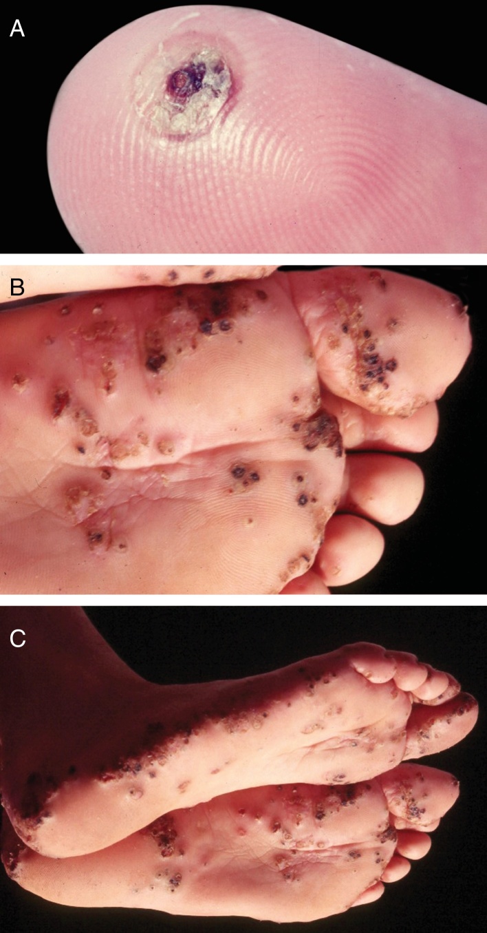
(A) Tungiasis – typical aspect. Isolated lesion. Note a pustule, in regression, with central crusted area. (B, C) Tungiasis. Multiple lesions, isolated and confluent.

Bedbug dermatitis. Multiple, characteristic linear erythematous papular lesions located in the abdomen.

Furunculoid myiasis. Ulcero-nodular lesion and etiological agent (Dermatobia hominis).

(A) Larva migrans. Intense pruritus. Typical serpiginous lesion with linear aspect. (B) Larva migrans. Numerous lesions caused by multiple larvae.

Lyme disease. Plaque presenting centrifugal growth, with erythematous-violet borders, measuring approximately 18 cm, located on the posterior surface of the thigh.

Onchocerciasis. Intense pruritus, presence of lichenification, exulcerations, and hyperpigmentation. Patient from the Infectious Disease Clinic, Ibadan, Nigeria (personal archive: Prof. Dr. Sinésio Talhari).

A. Onchocerciasis. Presence of atrophy, common in patients with long course. Native Brazilian from the Yanomami tribe (personal archive: Prof. Dr. Sinésio Talhari). B. Onchocerciasis. Observe the classic aspect of the “hanging groin” due to long evolution. There is also scrotum elongation (secondary to cutaneous atrophy), and there are nodules in the iliac crest and left groin – probably onchocercomas. Native Brazilian from the Yanomami tribe (personal archive: Prof. Dr. Sinésio Talhari).
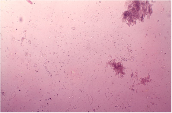
Onchocerciasis. Observe two microfilariae (Onchocerca volvulus). Direct examination in saline solution. 40× magnification (personal archive: Prof. Dr. Sinésio Talhari).
References
-
- Arlian L.G., Runyan R.A., Achar S., Estes S.A. Survival and infectivity of Sarcoptes scabiei var. canis and var. hominis. J Am Acad Dermatol. 1984;11:210–215. - PubMed
-
- Arlian lG, Estes S.A., Vyszenskimoher D.L. Prevalence of Sarcoptes scabiei in the homes and nursing homes of scabietic patients. J Am Acad Dermatol. 1988;19:806–811. - PubMed
-
- Rubegni P., Mandato F., Risulo M., Fimiani M. Non-invasive diagnosis of nodular scabies: the string of pearls sign. Australas J Dermatol. 2011;52:79. - PubMed
-
- Suh KS, Han SH, Lee KH, Park JB, Jung SM, Kim ST, et al. Mites and burrows are frequently found in nodular scabies by dermoscopy and histopathology. J Am Acad Dermatol. 2014;71:1022–3. - PubMed
-
- Cölgeçen-Özel E., Ertaş R., Utaş S., Kontaş O. Scabies mimicking mastocytosis in two infants. Turk J Pediatr. 2013;55:533–535. - PubMed
Publication types
MeSH terms
LinkOut - more resources
Full Text Sources
