Review
. 2020 Jun:102:113-121.
doi: 10.1016/j.semcdb.2019.12.003. Epub 2020 Jan 27.

Regulation of actin isoforms in cellular and developmental processes

Affiliations
Review

Regulation of actin isoforms in cellular and developmental processes

Anna S Kashina. Semin Cell Dev Biol. 2020 Jun.

Abstract

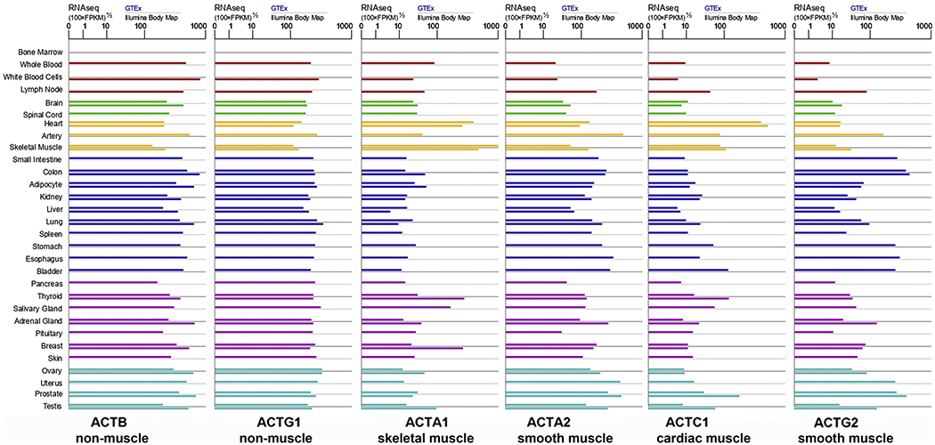
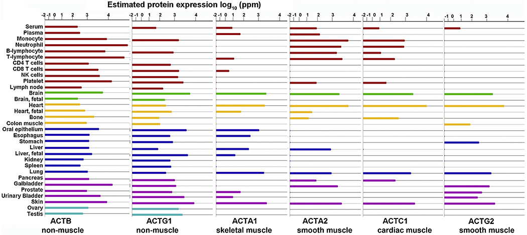
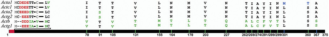
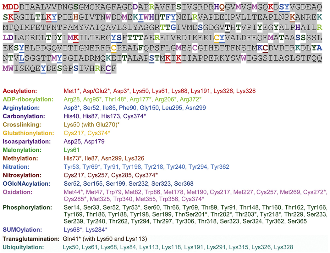
Actin is one of the most abundant and essential intracellular proteins that mediates nearly every form of cellular movement and underlies such key processes as embryogenesis, tissue integrity, cell division and contractility of all types of muscle and non-muscle cells. In mammals, actin is represented by six isoforms, which are encoded by different genes but produce proteins that are 95-99 % identical to each other. The six actin genes have vastly different functions in vivo, and the small amino acid differences between the proteins they encode are rigorously maintained through evolution, but the underlying differences behind this distinction, as well as the importance of specific amino acid sequences for each actin isoform, are not well understood. This review summarizes different levels of actin isoform-specific regulation in cellular and developmental processes, starting with the nuclear actin's role in transcription, and covering the gene-level, mRNA-level, and protein-level regulation, with a special focus on mammalian actins in non-muscle cells.

Keywords: Actin; Coding sequence; Posttranslational modifications; Regulation of cytoskeleton; Translation rate.


Figures

Figure 1.
Figure 1.. mRNAs for the six mammalian actin isoforms are abundant in many mammalian tissues.
Data was adapted and simplified from the charts available at https://www.genecards.org/ for the human actin genes.
Figure 2.
Figure 2.. Six mammalian actin isoforms show differential tissue abundance at the protein level.
Data was adapted and simplified from the charts available at https://www.genecards.org/ for the human actin genes.
Figure 3.
Figure 3.. Six mammalian actin isoforms are highly conserved at the amino acid level.
The solid line at the bottom represents the alignment of the six mammalian actins (following the gene symbols listed on the left). Red bar on the alignment represents the sequences unique to each actin isoform. Green bars represent the point amino acid substitutions that distinguish muscle from non-muscle actins. Blue represent the substitutions unique to only one actin isoform (a-skeletal actin). Letters on top list the amino acids in each of these positions for each isoforms (order corresponds to the gene list on the left). Dashes represent empty spaces. Numbers underneath represent the positions of these substitutions, counting from the initiator Met.
Figure 4.
Figure 4.. Actin undergoes a large variety of posttranslational modifications.
Top, amino acid sequence of mammalian β–actin, color-coded to depict sites of all modifications, listed alphabetically underneath. Underlined residues represent sites of multiple modifications, color-coded for the one found first in the list underneath. Areas shaded in gray represent the residues on which no posttranslational modifications have been described. Please note that amino acid positions in the list correspond to those reported in the published studies and are often counted not from the first Met but from the third residue, which appears as N-terminal after posttranslational processing in muscle actins. This list was adapted and updated from (Terman and Kashina, 2013), which lists many of the original publications that reported these arginylated sites.
Figure 5.
Figure 5.. Sites of actin’s posttranslational modifications mapped onto the folded actin monomer.
Yellowed residues represent the sites of possible posttranslational modifications, listed and mapped in Figure 3. Pink residues represent the sites where no posttranslational modifications have been described. The actin structure is based on the Cn3D view of b-actin (PDB identifier 1HLU).
Figure 6.
Figure 6.. Levels of intracellular actin regulation.
Actin is regulated at the gene level, through mRNA stability and accessibility to translation, as well as through interactions with multiple binding partners and direct targeting by posttranslational modifications. Interplay of this regulation dynamically controls the diversity of actin functions.

References

    1. A M, Fung TS, Kettenbach AN, Chakrabarti R, and Higgs HN (2019). A complex containing lysine-acetylated actin inhibits the formin INF2. Nature cell biology 21, 592–602. - PMC - PubMed
    1. Abe A, Saeki K, Yasunaga T, and Wakabayashi T (2000). Acetylation at the N-terminus of actin strengthens weak interaction between actin and myosin. Biochemical and biophysical research communications 268, 14–19. - PubMed
    1. Akimoto Y, Yan K, Miura Y, Tsumoto H, Toda T, Fukutomi T, Sugahara D, Kudo A, Arai T, Chiba Y, et al. (2019). O-GlcNAcylation and Phosphorylation of beta-Actin Serine199 in Diabetic Nephropathy. American journal of physiology Renal physiology. - PMC - PubMed
    1. Ampe C, and Van Troys M (2017). Mammalian Actins: Isoform-Specific Functions and Diseases. Handbook of experimental pharmacology 235, 1–37. - PubMed
    1. Anthony Akkari P, Nowak KJ, Beckman K, Walker KR, Schachat F, and Laing NG (2003). Production of human skeletal alpha-actin proteins by the baculovirus expression system. Biochemical and biophysical research communications 307, 74–79. - PubMed

Publication types

LinkOut - more resources