. 2020 Apr;10(4):552-567.
doi: 10.1158/2159-8290.CD-19-0813. Epub 2020 Jan 30.

Impaired Death Receptor Signaling in Leukemia Causes Antigen-Independent Resistance by Inducing CAR T-cell Dysfunction

Affiliations

Impaired Death Receptor Signaling in Leukemia Causes Antigen-Independent Resistance by Inducing CAR T-cell Dysfunction

Nathan Singh et al. Cancer Discov. 2020 Apr.

Abstract

Primary resistance to CD19-directed chimeric antigen receptor T-cell therapy (CART19) occurs in 10% to 20% of patients with acute lymphoblastic leukemia (ALL); however, the mechanisms of this resistance remain elusive. Using a genome-wide loss-of-function screen, we identified that impaired death receptor signaling in ALL led to rapidly progressive disease despite CART19 treatment. This was mediated by an inherent resistance to T-cell cytotoxicity that permitted antigen persistence and was subsequently magnified by the induction of CAR T-cell functional impairment. These findings were validated using samples from two CAR T-cell clinical trials in ALL, where we found that reduced expression of death receptor genes was associated with worse overall survival and reduced T-cell fitness. Our findings suggest that inherent dysregulation of death receptor signaling in ALL directly leads to CAR T-cell failure by impairing T-cell cytotoxicity and promoting progressive CAR T-cell dysfunction. SIGNIFICANCE: Resistance to CART19 is a significant barrier to efficacy in the treatment of B-cell malignancies. This work demonstrates that impaired death receptor signaling in tumor cells causes failed CART19 cytotoxicity and drives CART19 dysfunction, identifying a novel mechanism of antigen-independent resistance to CAR therapy.See related commentary by Green and Neelapu, p. 492.


Conflict of interest statement

Competing interests: C.H.J. has received grant support from Novartis, and has patents related to CAR therapy with royalties paid from Novartis to the University of Pennsylvania. C.H.J. is also a scientific founder and holds equity in Tmunity Therapeutics. S.A.G. has received support from Novartis, Servier and Kite, and serves as a consultant, member of the scientific advisory board or study steering committee for Novartis, Cellectis, Adaptimmune, Eureka, TCR2, Juno, GlaxoSmithKline, Vertex, Cure Genetics, Humanigen and Roche. N.S., S.G. and M.R. hold patents related to CAR T cell therapy. E.J.O. and K.T.M. are employed by Novartis. All other authors declare no relevant competing interests.

Figures

Figure 1.
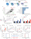
Figure 1.. Death receptor signaling is a key mediator of resistance to CAR T cell therapy.
(A) Schematic of genome-wide knockout screen of Brunello library-edited Nalm6 ALL cells and CART19. (B) Scatter plot of normalized MAGeCK beta scores representing enriched and depleted sgRNAs after Brunello screen. (C) Gene set enrichment analysis of the most-enriched and most-depleted sgRNAs identified after screen. (D) Survival over time of WT, FADDKO and BIDKO Nalm6 combined with CART19. (E) GFP+ Nalm6 FADDKO or (F) BIDKO cells were combined with GFP-negative WT Nalm6 (25% GFP+KO with 75% GFP-negative WT cells) and co-cultured with either control of CART19 cells. Proportion of GFP+ cells over time is shown. (G) GFP+ WT Nalm6 cells were combined with either control or CART19 cells in the presence of birinipant or vehicle and Nalm6 survival was measured over time. (H-J) Survival over time of WT, FADDKO and BIDKO (H) Nalm6 combined with CART22, (I) OCI-Ly10 combined with CART19, and (J) Nalm6 combined with an alternative CD19-targeted CAR bearing the CD28 co-stimulatory domain. (K-L) Survival of immunodeficient mice engrafted with (K) FADDKO or (L) BIDKO Nalm6 after treatment with control or CART19 cells. *P<0.05, **P<0.01, ***P<0.001, ****P<0.0001, ns: not significant
Figure 2.
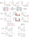
Figure 2.. Persistent exposure to ALL promotes progressive CART19 dysfunction.
(A) T cell expansion over time after combination with Nalm6. (B) Perforin and (C) granzyme B concentrations in culture supernatants over time after combination of CART19 and Nalm6 cells. (D) Schematic of functional evaluation studies, in which CART19 cells were sorted after short-term (5 day) or long-term (15 day) initial co-culture with either WT or BIDKO Nalm6, and then re-exposed to WT Nalm6 in secondary cultures. (E) CART19 expansion after 48h and 120h of secondary culture after short-term initial culture. (F) Cytokine secretion after 48h of secondary culture after short-term initial culture. (G) Nalm6 survival after 48h and 120h of secondary culture after short-term initial culture. (H) CART19 expansion after 48h and 120h of secondary culture after long-term initial culture. (I) Cytokine secretion after 48h of secondary culture after long-term initial culture. (J) Nalm6 survival after 48h and 120h of secondary culture after long-term initial culture. (K-L) CART19 cells were combined with WT or BIDKO Nalm6 cells either once at the beginning of co-culture or repeatedly and then sorted and re-cultured with WT Nalm6 as described. CART19 expansion was measured after 48h and 120h of secondary culture after (K) short-term and (L) long-term initial co-culture. (M-N) WT, PRF1KO or TRAILKO CART19 cells were combined with WT Nalm6 cells and sorted as described. CART19 expansion was measured after 48h and 120h of secondary culture after (M) short-term and (N) long-term initial co-culture. *P<0.05, **P<0.01, ***P<0.001, ****P<0.0001, ns: not significant
Figure 3.
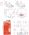
Figure 3.. Persistent exposure to Nalm6 drives molecular programs of dysfunction in CART19.
(A) Cell surface expression of PD-1 and (B) Tim3 over time on CART19 cells combined with WT or BIDKO Nalm6 either once at the beginning of culture or repeatedly. (C) Principal component analysis of gene expression profiles of CART19 cells at rest or after 15 days of co-culture with WT or BIDKO Nalm6. (D) Volcano plot of differentially-expressed genes in CART19 cells exposed to either WT or BIDKO Nalm6 for 15 days. Transcripts with log-fold change >0.5 and FDR <0.05 are shown in red. (E) Heatmap of differentially-accessible chromatin sites in CART19 cells at rest or after 15 days of co-culture with WT or BIDKO Nalm6. (F) Exemplary ATACseq tracks indicating increased chromatin accessibility (highlighted in grey) in T cell inhibitory transcription factors (noted in red) or decreased chromatin accessibility in pro-inflammatory transcription factor KLF2 (noted in green). *P<0.05, **P<0.01, ***P<0.001, ****P<0.0001, ns: not significant
Figure 4.
Figure 4.. Death receptor gene expression correlates with outcomes after tisagenlecleucel.
(A) Heatmap of RNA expression in pre-treatment leukemia-infiltrated bone marrow collected from patients treated with tisagenlecleucel (CR, n=18; NR, n=8). (B) Integrated gene set expression score of death receptor-associated pro-apoptotic genes (“death receptor signature”) in CRs and NRs from pediatric patients with ALL. High and low scores were defined using receiver operating curve analysis. (C) Overall survival of all pediatric patients analyzed. (D-E) Pharmacokinetic analysis of tisagenlecleucel in peripheral blood over the first (D) 90 days and (E) 1000 days after infusion, as measured by qPCR of CAR transcripts in patients with high (n=20) or low (n=6) death receptor signature scores. (F) Death receptor signature scores in CRs and NRs from adult patients with ALL. High and low scores were again defined by receiver operating curve analysis. (G) Overall survival of all adult patients analyzed.
Figure 5.
Figure 5.. Single cell analysis reveals the development of CAR T cell dysfunction in patients.
(A) UMAP projection of T cells contained in tisagenlecleucel infusion products prepared for pediatric patients who would go on to have either a complete response or no response after treatment. Cells are color-coded to differentiate the responding (grey) and non-responding (gold) patient. (B) T cells from the infusion products are color-coded to indicate expression of exhaustion-related gene transcripts (see Supplementary Table 3 for list of genes). (C) Bar graphs reflecting proportion of T cells from the infusion products that express 0, 1, 2, 3 or ≥4 exhaustion-related genes. Similar representations are shown for (D-F) peripheral blood T cells collected at peak expansion after tisagenlecleucel infusion, (G-I) CAR+ T cells in the infusion products and (J-L) peripheral blood CAR+ T cells collected at peak expansion after tisagenlecleucel infusion.
Figure 6.
Figure 6.
Proposed model for mechanism of impaired tumor death receptor signaling that leads to CAR T cell dysfunction and therapeutic failure.

Comment in

References

    1. Maude SL et al. Tisagenlecleucel in Children and Young Adults with B-Cell Lymphoblastic Leukemia. N Engl J Med 378, 439–448, doi: 10.1056/NEJMoa1709866 (2018). - DOI - PMC - PubMed
    1. Schuster SJ et al. Tisagenlecleucel in Adult Relapsed or Refractory Diffuse Large B-Cell Lymphoma. N Engl J Med 380, 45–56, doi: 10.1056/NEJMoa1804980 (2019). - DOI - PubMed
    1. Park JH et al. Long-Term Follow-up of CD19 CAR Therapy in Acute Lymphoblastic Leukemia. N Engl J Med 378, 449–459, doi: 10.1056/NEJMoa1709919 (2018). - DOI - PMC - PubMed
    1. Turtle CJ et al. Immunotherapy of non-Hodgkin's lymphoma with a defined ratio of CD8+ and CD4+ CD19-specific chimeric antigen receptor-modified T cells. Sci Transl Med 8, 355ra116, doi: 10.1126/scitranslmed.aaf8621 (2016). - DOI - PMC - PubMed
    1. Lee DW et al. T cells expressing CD19 chimeric antigen receptors for acute lymphoblastic leukaemia in children and young adults: a phase 1 dose-escalation trial. Lancet 385, 517–528, doi: 10.1016/S0140-6736(14)61403-3 (2015). - DOI - PMC - PubMed

Publication types

MeSH terms

Substances