. 2020 Mar 6;295(10):3064-3079.
doi: 10.1074/jbc.RA119.011185. Epub 2020 Jan 30.

The noncanonical small heat shock protein HSP-17 from Caenorhabditis elegans is a selective protein aggregase

Affiliations

The noncanonical small heat shock protein HSP-17 from Caenorhabditis elegans is a selective protein aggregase

Manuel Iburg et al. J Biol Chem. .

Abstract

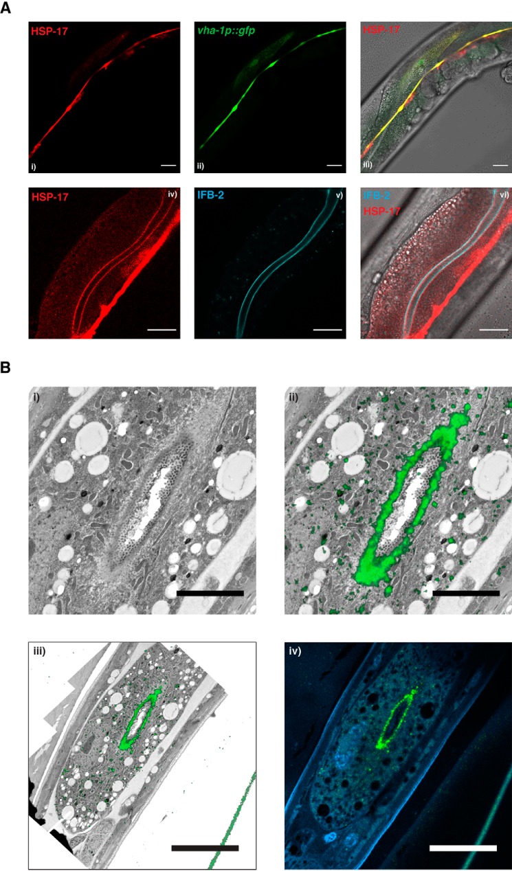
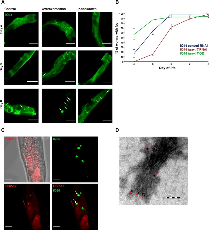
Small heat shock proteins (sHsps) are conserved, ubiquitous members of the proteostasis network. Canonically, they act as "holdases" and buffer unfolded or misfolded proteins against aggregation in an ATP-independent manner. Whereas bacteria and yeast each have only two sHsps in their genomes, this number is higher in metazoan genomes, suggesting a spatiotemporal and functional specialization in higher eukaryotes. Here, using recombinantly expressed and purified proteins, static light-scattering analysis, and disaggregation assays, we report that the noncanonical sHsp HSP-17 of Caenorhabditis elegans facilitates aggregation of model substrates, such as malate dehydrogenase (MDH), and inhibits disaggregation of luciferase in vitro Experiments with fluorescently tagged HSP-17 under the control of its endogenous promoter revealed that HSP-17 is expressed in the digestive and excretory organs, where its overexpression promotes the aggregation of polyQ proteins and of the endogenous kinase KIN-19. Systemic depletion of hsp-17 shortens C. elegans lifespan and severely reduces fecundity and survival upon prolonged heat stress. HSP-17 is an abundant protein exhibiting opposing chaperone activities on different substrates, indicating that it is a selective protein aggregase with physiological roles in development, digestion, and osmoregulation.

Keywords: Caenorhabditis elegans (C. elegans); chaperone; protein aggregates; protein aggregation; protein folding; proteostasis; selective protein aggregase; small heat shock protein (sHsp).


Conflict of interest statement

The authors declare that they have no conflicts of interest with the contents of this article

Figures

Figure 1.
Figure 1.
HSP-17 promotes aggregation of MDH. A, purification of HSP-17. Lanes 0–2, elution of protein from His6-NiNTA beads; lane R/C, removal of tag and rebuffering; lane I, filtered, diluted input for anion-exchange chromatography after removal of tag with His6-NiNTA beads; lane 17, fraction from anion-exchange chromatography containing HSP-17. B, SEC analysis of purified HSP-17 at a concentration of 1.5 mg/ml. Absorption at 220 nm, given in arbitrary units (au), shows a molecular mass of >440 kDa corresponding to a 26-mer. EV, exclusion volume; F1, start of first fraction for SDS-PAGE analysis; FL, start of last fraction sampled for SDS-PAGE analysis. Exclusion volumes of molecular weight standards are indicated at the top. C, MDH was aggregated at 41 °C (blue) and in the presence of increasing concentrations of HSP-17 (red shades). A darker hue of red indicates higher concentration of HSP-17. Light scattering was measured at 360 nm. Buffer alone (gray) and the highest concentration of HSP-17 in the absence of MDH (black) were plotted. D, as in C, at 47 °C. E, MDH was aggregated in the presence or absence of a 2-fold excess of HSP-17, at 41 or 47 °C. Samples were collected and centrifuged at 20,800 × g for 20 min to separate soluble (S) and pellet (P) fractions prior to SDS-PAGE analysis. The relative signal intensities of MDH and HSP-17 in the soluble and pellet fractions are listed in the table below. F, as in E, but the experiment was performed with HSP-17 alone, in the absence of MDH.
Figure 2.
Figure 2.
HSP-17 inhibits disaggregation and refolding of aggregated luciferase. A, CS was aggregated at 43 °C (blue) and in the presence of increasing concentrations of HSP-17 (red shades). A darker hue of red indicates higher concentration of HSP-17. Light scattering was measured at 360 nm. Buffer alone (gray) and the highest concentration of HSP-17 in absence of CS (black) were plotted. B, as in A, but with GAPDH as substrate instead of CS, at a temperature of 45 °C. C, schematic illustration of the experimental procedure of the assay carried out in D and E. I, aggregation of luciferase in the absence of HSP-17. II, aggregation of luciferase in presence of HSP-17. D, firefly luciferase was subjected to heat shock to form aggregates that were subsequently resolubilized and refolded by HSP-1, DNJ-12, DNJ-13, and HSP-110. Recovery is measured over 2 h by a regain of its enzymatic function via luminescence signal normalized to native luciferase (blue). Varying concentrations of HSP-17 have been added during heat-mediated aggregation of luciferase (darker hues of red indicating higher concentrations of HSP-17). E, as in C, but HSP-17 was added after aggregation of luciferase.
Figure 3.
Figure 3.
HSP-17 is expressed in the alimentary system and the excretory canal. A, confocal imaging of the translational HSP-17 reporter phsp-17::hsp-17::wrmScarlet transgene. Left, starting from the bottom, the arrow points to pharynx, excretory canal cell, vulva, and anus. Scale bar, 100 μm. i, localization to the pharynx. Scale bar, 20 μm. ii, localization to the intestine and particularly the membrane around the lumen (arrow). Scale bar, 20 μm. iii, left arrow, cells underneath the cuticle; right arrow, pseudocoelom around an egg in utero. Scale bar, 50 μm. iv, localization to anus. Scale bar, 20 μm. v, a spotted line of HSP-17::wrmScarlet around one of the excretory canals (arrow; vesicular activity). Scale bar, 50 μm. vi, localization to vulva (arrow). Scale bar, 50 μm. B, Western blotting of C. elegans populations of N2, HSP-17::GFP, and HSP-17::wrmScarlet animals synchronized to 4 days of age. Endogenous HSP-17 and the fusion proteins with the fluorophores were detected using polyclonal HSP-17 antibodies. Tubulin was used for normalization of signal intensities. The relative intensities for the endogenous and fluorescently tagged HSP-17 are provided below the blot. C, lifespan of hsp-17-depleted N2 (hsp-17 RNAi; red) and control animals (empty vector; blue). 150 animals were analyzed for each condition. The mean lifespan is indicated in the inset. Maximum lifespans are 29 (control)/27 (RNAi) days, respectively. Log-rank test yields p = 0.0003. D, fecundity assay of RNAi control, hsp-17 RNAi, WT N2, and transgenic HSP-17 overexpression lines. Depicted are the numbers of offspring. Error bars, S.D. Triplicates of 20 animals were analyzed for each condition. E, RNAi control, hsp-17 RNAi, and the HSP-17 transgenic animals were subjected to a 6-h heat shock of 35 °C before allowing them to recover at 20 °C for 24 h. The graph depicts the percentage of survivors. Error bars, S.D. Triplicates of at least 50 animals were analyzed for each transgenic condition. F, C. elegans N2 populations were synchronized to ages of 2, 4, 6, and 8 days, and their lysates were separated into soluble and insoluble fractions for subsequent detection of HSP-17 by Western blotting. The graph depicts the relative intensities of HSP-17 in each fraction. S.E. is given. G, 4-day-old nematodes of WT N2, N2 + hsp-17 RNAi, and hsp-17 OE animals were transferred to NGM plates containing 250 mm NaCl. Survivors were counted until all nematodes died. 20 animals/condition were analyzed in three replicates. *, p < 0.05; **, p < 0.01; ****, p < 0.0001.
Figure 4.
Figure 4.
HSP-17 is localized to the excretory tract and to the intestinal endotube. A, translational fluorescent reporters of hsp-17 were crossed with reporter strains for visualization of in vivo localization and interactions. The red (560-nm) channel for wrmScarlet fluorescence is on the left, the GFP or CFP channels in the middle, and the overlay of both fluorescent channels and brightfield on the right. i–iii, co-localization of Pvha-1::gfp and Phsp-17::hsp-17::wrmScarlet; iv–vi, co-localization of intestinal intermediary filament B-2 (ifb-2::cfp) and Phsp-17::hsp-17::wrmScarlet. Scale bars, 20 μm. B, Bottom: total perspective of the CLEM-image in i and ii. Left side (iii) shows an overlay of fluorescence image and electron microscopy, right side (iv) includes the DAPI channel. Scale bars are 4 μm. (i and ii) or 10 μm (iii and iv).
Figure 5.
Figure 5.
HSP-17 promotes formation of polyQ foci in vivo. A, C. elegans expressing Q44::YFP in the intestine (iQ44) were monitored from day 4 to day 8 of life, and nematodes with polyQ foci were quantified. iQ44 nematodes were subjected to hsp-17 RNAi and RNAi control conditions. In addition, iQ44 nematodes overexpressing the HSP-17::wrmScarlet fusion were analyzed. Depicted are representative images of the data on days 4, 5, and 6 of life. White arrows, polyQ foci. Scale bars, 50 μm. B, the percentage of nematodes showing foci in A was plotted over days of life. Error bars, S.D.; 20 animals/condition were analyzed in duplicates. C, analysis of the co-localization of HSP-17 (HSP-17::wrmScarlet) with intestinal Q85-YFP. Depicted are the individual channels as well as the overlay. White arrows, foci of co-localization between HSP-17-wrmScarlet and iQ85-YFP. Scale bars, 20 μm. D, interaction of purified HSP-17 with HttEx1Q48 fibrils analyzed using HSP-17 antibodies. Immunogold signals depicting the association of HSP-17 with the HttEx1Q48 fibrils are indicated by red triangles. The image shows 85,000-fold magnification; scale bar, 100 nm.
Figure 6.
Figure 6.
HSP-17 overexpression leads to increased aggregation of KIN-19. A, fluorescent images of the localization and foci formation of KIN-19-RFP (CF3166) and co-localization with HSP-17::GFP. i and iv, individual channels (488 nm) of HSP-17::GFP; ii and v, individual channels (560 nm) of KIN-19-RFP. i and ii, excretory canal; iv and v, pharynx; iii and vi, overlays of both fluorescent channels and bright field. The co-localization of KIN-19 foci with HSP-17 is particularly evident in the pharynx (vii). Scale bars, 20 μm. B, quantification of KIN-19 foci in RNAi control, hsp-17 RNAi, and HSP-17 OE animals at the age of 5 days. 20 animals/condition were analyzed. C, developmental analysis of N2 or N2 subjected to control RNAi (dark blue), N2 subjected to hsp-17 RNAi (red), HSP-17 overexpression (green), KIN-19 overexpression or KIN-19 overexpression subjected to control RNAi (purple), HSP-17 and KIN-19 overexpression (light blue), and KIN-19 overexpression subjected to RNAi of hsp-17 (orange). Depicted are the percentages of animals that reach adulthood 4 days after hatching at 20 °C. *, p < 0.05; **, p < 0.01; ***, p < 0.001.

References

    1. Hartl F. U., Bracher A., and Hayer-Hartl M. (2011) Molecular chaperones in protein folding and proteostasis. Nature 475, 324–332 10.1038/nature10317 - DOI - PubMed
    1. Morimoto R. I. (2008) Proteotoxic stress and inducible chaperone networks in neurodegenerative disease and aging. Genes Dev. 22, 1427–1438 10.1101/gad.1657108 - DOI - PMC - PubMed
    1. Richter K., Haslbeck M., and Buchner J. (2010) The heat shock response: life on the verge of death. Mol. Cell 40, 253–266 10.1016/j.molcel.2010.10.006 - DOI - PubMed
    1. Tyedmers J., Mogk A., and Bukau B. (2010) Cellular strategies for controlling protein aggregation. Nat. Rev. Mol. Cell Biol. 11, 777–788 10.1038/nrm2993 - DOI - PubMed
    1. Chiti F., and Dobson C. M. (2017) Protein misfolding, amyloid formation, and human disease: a summary of progress over the last decade. Annu. Rev. Biochem. 86, 27–68 10.1146/annurev-biochem-061516-045115 - DOI - PubMed

Publication types

MeSH terms