. 2020 Mar 13;295(11):3692-3707.
doi: 10.1074/jbc.RA119.010497. Epub 2020 Jan 30.

The DNA repair enzyme MUTYH potentiates cytotoxicity of the alkylating agent MNNG by interacting with abasic sites

Affiliations

The DNA repair enzyme MUTYH potentiates cytotoxicity of the alkylating agent MNNG by interacting with abasic sites

Alan G Raetz et al. J Biol Chem. .

Abstract

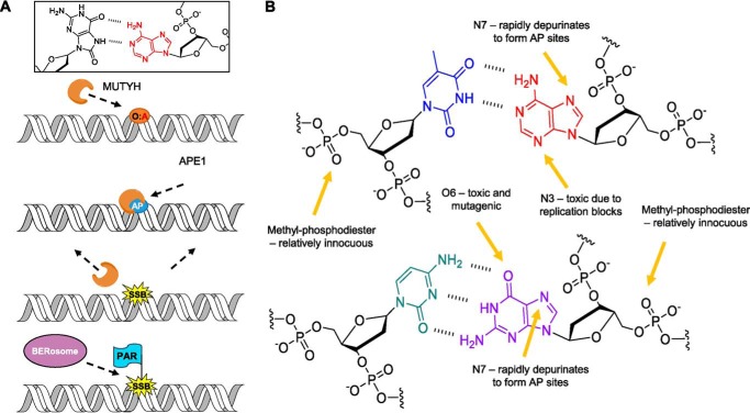
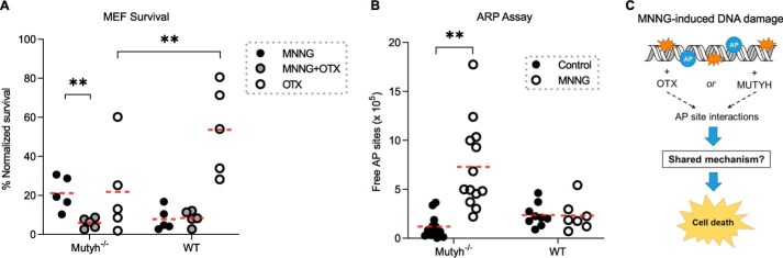
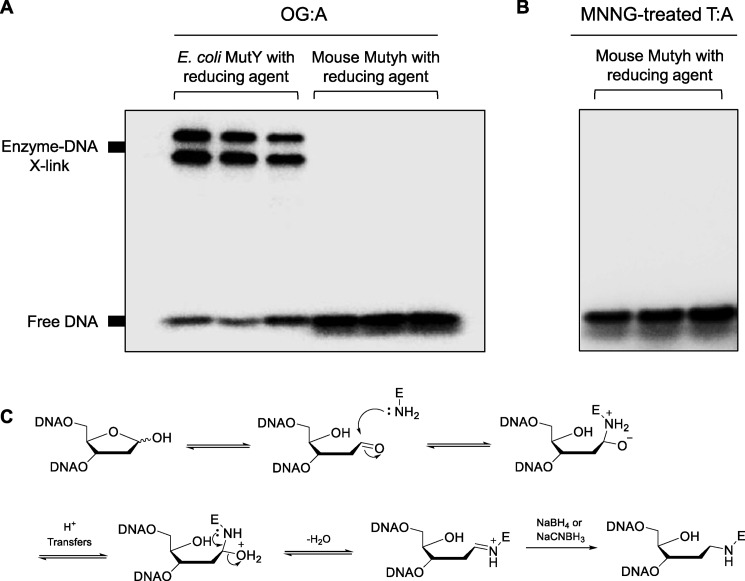
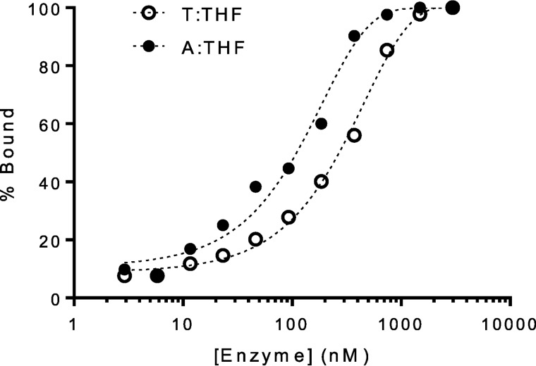
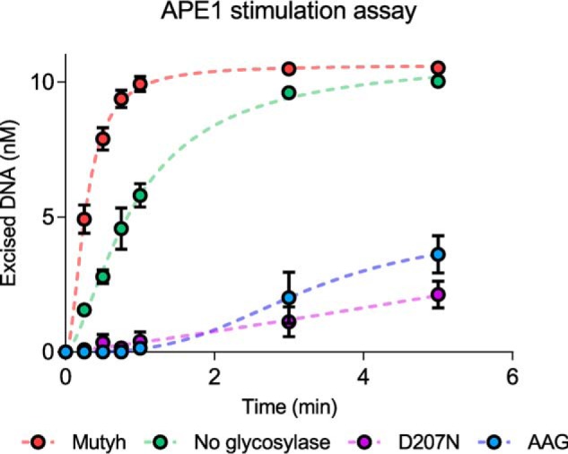
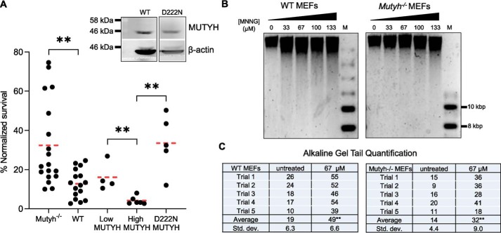
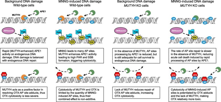
Higher expression of the human DNA repair enzyme MUTYH has previously been shown to be strongly associated with reduced survival in a panel of 24 human lymphoblastoid cell lines exposed to the alkylating agent N-methyl-N'-nitro-N-nitrosoguanidine (MNNG). The molecular mechanism of MUTYH-enhanced MNNG cytotoxicity is unclear, because MUTYH has a well-established role in the repair of oxidative DNA lesions. Here, we show in mouse embryonic fibroblasts (MEFs) that this MNNG-dependent phenotype does not involve oxidative DNA damage and occurs independently of both O6-methyl guanine adduct cytotoxicity and MUTYH-dependent glycosylase activity. We found that blocking of abasic (AP) sites abolishes higher survival of Mutyh-deficient (Mutyh-/-) MEFs, but this blockade had no additive cytotoxicity in WT MEFs, suggesting the cytotoxicity is due to MUTYH interactions with MNNG-induced AP sites. We found that recombinant mouse MUTYH tightly binds AP sites opposite all four canonical undamaged bases and stimulated apurinic/apyrimidinic endonuclease 1 (APE1)-mediated DNA incision. Consistent with these observations, we found that stable expression of WT, but not catalytically-inactive MUTYH, enhances MNNG cytotoxicity in Mutyh-/- MEFs and that MUTYH expression enhances MNNG-induced genomic strand breaks. Taken together, these results suggest that MUTYH enhances the rapid accumulation of AP-site intermediates by interacting with APE1, implicating MUTYH as a factor that modulates the delicate process of base-excision repair independently of its glycosylase activity.

Keywords: DNA damage; DNA damage response; DNA enzyme; DNA methylation; DNA repair; abasic sites; base excision repair (BER); cancer biology; cancer chemoprevention; mutY homolog (MUTYH).


Conflict of interest statement

The authors declare that they have no conflicts of interest with the contents of this article

Figures

Figure 1.
Figure 1.
Oxidative and alkylative base damage and repair. A, depiction of MUTYH's known canonical role in the repair of OG:A (O:A) lesions in the cell, followed by downstream processing by APE1 and other BER proteins. B, functional groups within dsDNA that are susceptible to methylation by MNNG. Thymine (blue) is paired with adenine (red), and cytosine (teal) is paired with guanine (purple).
Figure 2.
Figure 2.
MUTYH expression alters MEF survival to MNNG. A, normalized survival versus control condition 6–7 days after treating with low MNNG (67 μm) in Mutyh−/−, wildtype (WT), and Mutyh−/− MEFs stable cell lines expressing recombinant human MUTYH, classified as low MUTYH (expression) and high MUTYH (expression) based on Western blotting. Data are from at least four biological replicates. **, p < 0.005, t test, significant with Bonferroni correction for multiple comparisons. B, Western blotting of representative Mutyh−/− MEF stable cell lines transfected with the pcDNA3.1 human MUTYH construct after G418 antibiotic selection. HEK-293 cell lysate is shown as a MUTYH-positive control. Clones A and C were categorized as high MUTYH expression. Clones B and D were categorized as low MUTYH expression. The figure is composed of two separate Western blottings using the same antibodies and conditions as detailed under “Experimental procedures.”
Figure 3.
Figure 3.
Under conditions of high MNNG, which induce oxidative stress, Mutyh enhances cell survival. A and B, ROS detection using the redox-sensitive fluorescent probe H2-DCFDA in MNNG-treated WT MEFs (A) versus hydrogen peroxide-treated WT MEFs as a positive control (B). Each point represents the percent of fluorescence-positive cells from an independent flow cytometry experiment as detailed under “Experimental procedures.” The trend line represents the mean percent fluorescent cells as a function of concentration. C, 6–7-day survival of WT and Mutyh−/− MEFs at high concentration MNNG from four separate experiments (averaged data from combined 333 and 667 μm treatments). WT MEFs had significantly higher survival under these conditions (**p = 0.01). D, survival of Ogg1−/− MEFs to 67 μm MNNG is not significantly different from their matched parental WT MEFs (p = 0.64). E, detection of MNNG-induced lesions by purified Ogg1 enzyme plasmid nicking assay, where the increased density of the upper open circular (oc) DNA indicates strand nicking versus the closed-circular (cc) plasmid DNA.
Figure 4.
Figure 4.
Recombinant Mutyh does not possess methyl-base glycosylase activity. The 1st five panels are representative LC traces for each condition (HCl, no enzyme (No Enz, negative control), AAG (positive control), WT Mutyh, and D207N Mutyh), with signal intensity monitored by MS counts. The last panel is a bar graph summarizing the detection of methylated bases quantified by LC-MS/MS for each experimental condition. All reactions were performed in experimental triplicates, and error bars represent the standard deviation from the average.
Figure 5.
Figure 5.
Evidence of AP site interactions in MUTYH-mediated MNNG toxicity. A, cell survival data of WT and Mutyh−/− MEFs treated with 33 μm MNNG with and without 3 mm OTX or with OTX alone. The enhanced survival of Mutyh−/− MEFs to MNNG is abolished by OTX (1st column versus 2nd column, p = 0.005) but has no effect on WT MEFs (4th column versus 5th column). OTX alone is significantly more toxic to Mutyh−/− versus WT MEFs (3rd column versus 6th column, p = 0.011). B, ARP assay quantification of AP sites in genomic DNA extracted from MEFs treated with MNNG versus untreated control. There was a significant increase in reactive AP sites in Mutyh−/− MEFs (1st column versus 2nd column) but not in WT MEFs upon MNNG treatment (p < 0.001, t test). C, diagram summarizing experimental results, which highlights the lack of an additive effect between the small molecule OTX and MUTYH, suggesting they potentiate MNNG cell death by a similar mechanism.
Figure 6.
Figure 6.
Trapping of transient Mutyh–DNA complexes with reducing agents. In vitro cross-linking assays with bacterial and mammalian MutY enzymes with OG:A-containing 30-bp DNA (A) or MNNG-treated T:A-containing 30-bp DNA (B) using 90 mm of either NaBH4 or NaCNBH3 as reducing agents. A, 1st and 2nd lanes, DNA with E. coli MutY and NaBH4; 3rd lane, DNA E. coli MutY and NaCNBH3; 4th to 6th lanes are the same conditions as the 1st to 3rd lanes, but with WT mouse Mutyh. B, 1st lane, no enzyme control; 2nd lane, DNA with mouse Mutyh; 3rd lane, DNA with mouse MUTYH and NaBH4. C, proposed mechanism for formation of a transient Schiff base intermediate between MutY and substrate DNA, followed by reductive “trapping” of the enzyme–DNA complex.
Figure 7.
Figure 7.
Mutyh exhibits high affinity for AP sites. Representative fluorescence polarization data used for apparent Kd determination with Mutyh and X:THF-containing DNA, where X = T or A. Table 1 lists the experimentally determined apparent Kd values for a given base pair context.
Figure 8.
Figure 8.
Mutyh stimulates APE1 activity. Representative data for in vitro APE1 stimulation assays performed with either glycosylase-free or with glycosylase-bound duplex DNA containing a centrally-located T:THF lesion. Reactions were conducted at 37 °C for 40 min under multiple turnover conditions and quenched by the addition of 0.2 m NaOH at preselected time points, as described under “Experimental procedures.” Dashed lines are included for illustration purposes, and do not represent fits to experimental data.
Figure 9.
Figure 9.
MNNG-dependent cytotoxicity depends on MUTYH catalytic residue Asp-222 and leads to genomic DNA strand breaks. A, survival to low MNNG (67 μm) in Mutyh−/− MEF stable cell lines expressing recombinant D222N (5th column) versus WT MUTYH (4th column). Data from Fig. 2A are represented in the 1st to 4th columns. Inset: Western blotting of WT MUTYH expression from Fig. 2B versus D222N stable cell line MUTYH expression. There is significantly higher survival in cells expressing D222N versus WT MUTYH (4th column versus 5th column, **, p < 0.005, t test, significant with Bonferroni correction for multiple comparisons). B, MUTYH increases genomic strand breaks in MNNG-treated MEFs as quantified by alkaline gel electrophoresis. Genomic DNA was extracted 40 min after MNNG treatment, quantified, incubated in an alkaline loading buffer, and run on a 0.8% agarose gel overnight at pH 12.4 as detailed under “Experimental procedures.” M = molecular weight marker. C, quantification of total sample band density in the lower region (tail) versus the upper band from two biological replicates (five gels). WT cells had significantly more DNA in the lower region at 67 μm versus Mutyh−/− cells (**, t test, p < 0.01), indicative of increased strand breaks in WT versus Mutyh−/− MEFs. Trial 5 is shown, and all other gels and data are shown in Fig. S5.
Figure 10.
Figure 10.
Proposed model of MUTYH-mediated enhancement of MNNG cytotoxicity, with and without OTX.

References

    1. Al-Tassan N., Chmiel N. H., Maynard J., Fleming N., Livingston A. L., Williams G. T., Hodges A. K., Davies D. R., David S. S., Sampson J. R., and Cheadle J. P. (2002) Inherited variants of MYH associated with somatic G:C→T:A mutations in colorectal tumors. Nat. Genet. 30, 227–232 10.1038/ng828 - DOI - PubMed
    1. Cheadle J. P., and Sampson J. R. (2007) MUTYH-associated polyposis–from defect in base excision repair to clinical genetic testing. DNA Repair 6, 274–279 10.1016/j.dnarep.2006.11.001 - DOI - PubMed
    1. David S. S., O'Shea V. L., and Kundu S. (2007) Base-excision repair of oxidative DNA damage. Nature 447, 941–950 10.1038/nature05978 - DOI - PMC - PubMed
    1. Banda D. M., Nuñez N. N., Burnside M. A., Bradshaw K. M., and David S. S. (2017) Repair of 8-oxoG:A mismatches by the MUTYH glycosylase: mechanism, metals and medicine. Free Radic. Biol. Med. 107, 202–215 10.1016/j.freeradbiomed.2017.01.008 - DOI - PMC - PubMed
    1. Markkanen E., Dorn J., and Hübscher U. (2013) MUTYH DNA glycosylase: the rationale for removing undamaged bases from the DNA. Front. Genet. 4, 18 10.3389/fgene.2013.00018 - DOI - PMC - PubMed

Publication types

MeSH terms