An official website of the United States government
The .gov means it’s official.
Federal government websites often end in .gov or .mil. Before
sharing sensitive information, make sure you’re on a federal
government site.
The site is secure.
The https:// ensures that you are connecting to the
official website and that any information you provide is encrypted
and transmitted securely.
Cancer immunotherapies that target the programmed cell death 1 (PD-1):programmed death-ligand 1 (PD-L1) immune checkpoint pathway have ushered in the modern oncology era. Drugs that block PD-1 or PD-L1 facilitate endogenous antitumor immunity and, because of their broad activity spectrum, have been regarded as a common denominator for cancer therapy. Nevertheless, many advanced tumors demonstrate de novo or acquired treatment resistance, and ongoing research efforts are focused on improving patient outcomes. Using anti-PD-1 or anti-PD-L1 treatment against earlier stages of cancer is hypothesized to be one such solution. This Review focuses on the development of neoadjuvant (presurgical) immunotherapy in the era of PD-1 pathway blockade, highlighting particular considerations for biological mechanisms, clinical trial design, and pathologic response assessments. Findings from neoadjuvant immunotherapy studies may reveal pathways, mechanisms, and molecules that can be cotargeted in new treatment combinations to increase anti-PD-1 and anti-PD-L1 efficacy.
Competing interests: D.M.P. and S.L.T. report stock and other ownership interests from Aduro Biotech, Compugen, DNAtrix, Dragonfly Therapeutics, Ervaxx, Five Prime Therapeutics, FLX Bio, Jounce Therapeutics, Potenza Therapeutics, Tizona Therapeutics, and WindMIL; consulting or advisory role with AbbVie, Amgen, Bayer, Compugen, DNAtrix, Dragonfly Therapeutics, Dynavax, Ervaxx, Five Prime Therapeutics, FLX Bio, Immunocore, lmmunomic Therapeutics, Janssen Oncology, Medlmmune, Merck, Tizona Therapeutics, and Wind MIL; research funding from Bristol-Myers Squibb, Compugen, and Potenza Therapeutics; patents, royalties, and other intellectual property from Aduro Biotech, Bristol-Myers Squibb, and lmmunonomic Therapeutics; and travel, accommodations, and expenses from Bristol-Myers Squibb and Five Prime Therapeutics. J.M.T. reports receiving equipment and reagents from Akoya Biosciences; grants and personal fees from Bristol-Myers Squibb; and personal fees from Merck, Astra Zeneca, and Amgen.
Figures
Fig. 1.. Two potential mechanisms for the…
Fig. 1.. Two potential mechanisms for the enhancement of systemic antitumor T cell immunity after…
Fig. 1.. Two potential mechanisms for the enhancement of systemic antitumor T cell immunity after neoadjuvant PD-1 blockade.
PD-1 blockade could result in the “in situ” expansion of tumor-specific T cell clones already within the tumor microenvironment (left). This expansion and activation is largely driven by PD-L1– and PD-L2–expressing dendritic cells in the tumor. Tumor-specific tumor-infiltrating lymphocytes (TILs) may represent naïve T cells or T cells that have already been “primed” to tumor antigen (84) before PD-1 pathway blockade. In addition, tumor antigen–containing DCs originating in the tumor pick up tumor antigens and traffic to the tumor-draining lymph nodes, where they present antigens either ineffectively or in a tolerogenic fashion to tumor-specific T cells. PD-1 blockade could act at this point, enhancing productive stimulation of tumor-specific T cells or partially reversing tolerance induction. Activated T cells enter the circulation by way of efferent lymphatics and then egress into tissues.
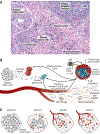
Fig. 2.. Immune-mediated tumor regression.
( A …
Fig. 2.. Immune-mediated tumor regression.
( A ) Representative photomicrograph from a definitive surgical resection…
Fig. 2.. Immune-mediated tumor regression.
(A) Representative photomicrograph from a definitive surgical resection specimen from a patient with NSCLC responding to neoadjuvant immunotherapy. Hematoxylin and eosin staining was used, with 100× original magnification. HEV, high endothelial venule. (B) Schematic of the tumor bed immunoarchitecture, displaying features consistent with both T cell– and B cell–mediated local antitumor immune responses. The regression bed—the area where the tumor used to be—is characterized by hallmarks of tissue repair and wound healing, such as neovascularization and proliferative fibrosis.(C) Percent RVT using irPRC is calculated by the surface area of the RVT/surface area of the tumor bed. The tumor bed surface area includes RVT + tumor-associated stroma + necrosis + regression bed. Schematics show examples of 100, 50, 10 (MPR), and 0% (pCR) RVT.
Topalian SL, Drake CG, Pardoll DM, Immune checkpoint blockade: A common denominator approach to cancer therapy. Cancer Cell 27, 450–461 (2015). doi: 10.1016/j.ccell.2015.03.001;
-
DOI
-
PMC
-
PubMed
Yarchoan M, Hopkins A, Jaffee EM, Tumor mutational burden and response rate to PD-1 inhibition. N. Engl. J. Med 377, 2500–2501 (2017). doi: 10.1056/NEJMc1713444
-
DOI
-
PMC
-
PubMed
Topalian SL et al., Safety, activity, and immune correlates of anti-PD-1 antibody in cancer. N. Engl. J. Med 366, 2443–2454 (2012). doi: 10.1056/NEJMoa1200690;
-
DOI
-
PMC
-
PubMed
Nghiem PT et al., PD-1 blockade with pembrolizumab in advanced Merkel-cell carcinoma. N. Engl. J. Med 374, 2542–2552 (2016). doi: 10.1056/NEJMoa1603702;
-
DOI
-
PMC
-
PubMed
Topalian SL, Taube JM, Anders RA, Pardoll DM, Mechanism-driven biomarkers to guide immune checkpoint blockade in cancer therapy. Nat. Rev. Cancer 16, 275–287 (2016). doi: 10.1038/nrc.2016.36;
-
DOI
-
PMC
-
PubMed