

Case Reports
doi: 10.1016/j.jcot.2019.06.014.
Epub 2019 Jun 14.
Proximal femoral fracture in ankylosed hip treated with primary total hip arthroplasty: Technical tips with report of two cases
Affiliations
- PMID: 32001994
- PMCID: PMC6985166
- DOI: 10.1016/j.jcot.2019.06.014
Item in Clipboard
Case Reports
Proximal femoral fracture in ankylosed hip treated with primary total hip arthroplasty: Technical tips with report of two cases
J Clin Orthop Trauma.
2020 Jan-Feb.
Erratum in
-
Erratum regarding missing declaration of competing interest statements in previously published articles.J Clin Orthop Trauma. 2020 Nov-Dec;11(6):1175. doi: 10.1016/j.jcot.2020.10.023. Epub 2020 Oct 15. J Clin Orthop Trauma. 2020. PMID: 33192026 Free PMC article.
-
Erratum regarding previously published articles.J Clin Orthop Trauma. 2021 Aug 5;21:101556. doi: 10.1016/j.jcot.2021.101556. eCollection 2021 Oct. J Clin Orthop Trauma. 2021. PMID: 34414070 Free PMC article.
Abstract
Proximal femoral fracture in an ankylosed hip is a challenging condition. There is no consensus on fixation method for these fractures. In addition, despite union the best outcome possible is the restoration of the pre morbid status. We report two different presentations of proximal femoral fracture in ankylosed hip that were successfully treated with primary total hip arthroplasty. We also discuss the surgical principles, technique and advantages of doing primary total hip arthroplasty in such cases.
© 2019.
Figures

a - b. Radiograph showing failed osteosynthesis for proximal femoral fracture in an arthrodesed hip in antero-posterior view (a) lateral view (b). Note the breakage of implant as well, which implies excessive stress over the fracture site due to ipsilateral fused hip. c - e. Serial radiographs after total hip arthroplasty at immediate post-operative period (a) at one month (b) at 6 months (c). Note the stable total hip prosthesis in situ showing total hip arthroplasty prosthesis in situ and healing of the fracture.
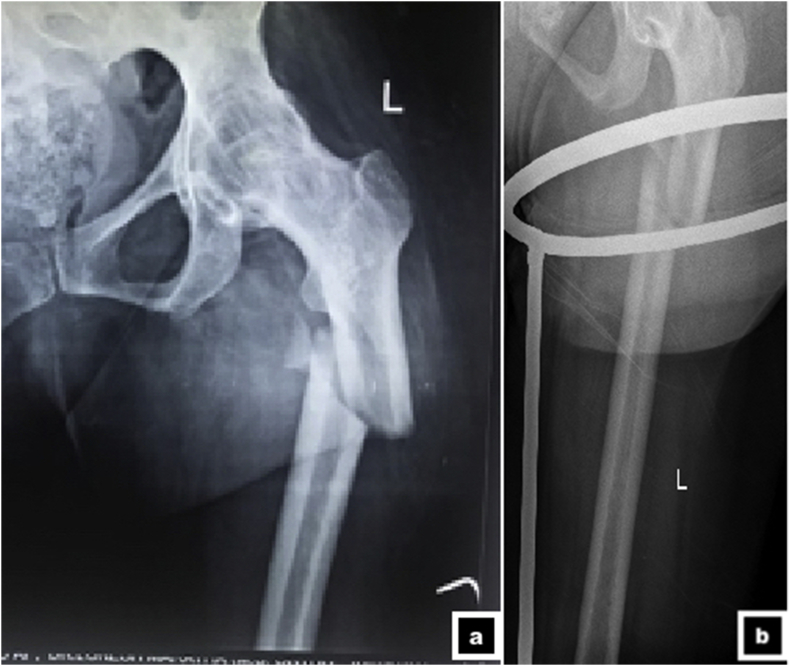
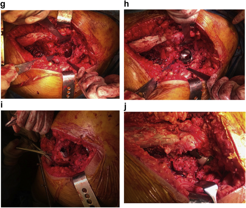
a - b. Radiograph showing sub-trochanteric fracture in an ankylosed left hip in antero-posterior view (a) oblique view (b). 2 c. Intra-operative picture showing posterior approach to the hip joint with exposure of proximal femur and femoral neck. Note the narrow access to the neck due to ankylosed hip. d. Intra-operative picture showing insitu femoral neck osteotomy. e. Intra-operative picture showing femoral preparation. The fracture is being provisionally stabilized over the canal reamer. f. Intra-operative picture showing acetabular preparation. The first reamer should be the smallest reamer to identify the floor of the acetabulum. g. Intra-operative picture showing the finally prepared acetabular bed prior to final cup placement. h Intra-operative picture showing final placement of acetabular cup. I Intra-operative picture showing placement of the final sleeve of a modular prosthesis (SROM®, Depuy Orthopedics, Warsaw, IN). j Intra-operative picture showing final reduction of the joint prior to closure. k - m. Serial radiographs after acute total hip arthroplasty at 6 weeks (a) at 3 months (b), and at 6 months (c). Note the stable total hip prosthesis in situ and union of the fracture.

a - b. Radiograph showing sub-trochanteric fracture in an ankylosed left hip in antero-posterior view (a) oblique view (b). 2 c. Intra-operative picture showing posterior approach to the hip joint with exposure of proximal femur and femoral neck. Note the narrow access to the neck due to ankylosed hip. d. Intra-operative picture showing insitu femoral neck osteotomy. e. Intra-operative picture showing femoral preparation. The fracture is being provisionally stabilized over the canal reamer. f. Intra-operative picture showing acetabular preparation. The first reamer should be the smallest reamer to identify the floor of the acetabulum. g. Intra-operative picture showing the finally prepared acetabular bed prior to final cup placement. h Intra-operative picture showing final placement of acetabular cup. I Intra-operative picture showing placement of the final sleeve of a modular prosthesis (SROM®, Depuy Orthopedics, Warsaw, IN). j Intra-operative picture showing final reduction of the joint prior to closure. k - m. Serial radiographs after acute total hip arthroplasty at 6 weeks (a) at 3 months (b), and at 6 months (c). Note the stable total hip prosthesis in situ and union of the fracture.
a - b. Radiograph showing sub-trochanteric fracture in an ankylosed left hip in antero-posterior view (a) oblique view (b). 2 c. Intra-operative picture showing posterior approach to the hip joint with exposure of proximal femur and femoral neck. Note the narrow access to the neck due to ankylosed hip. d. Intra-operative picture showing insitu femoral neck osteotomy. e. Intra-operative picture showing femoral preparation. The fracture is being provisionally stabilized over the canal reamer. f. Intra-operative picture showing acetabular preparation. The first reamer should be the smallest reamer to identify the floor of the acetabulum. g. Intra-operative picture showing the finally prepared acetabular bed prior to final cup placement. h Intra-operative picture showing final placement of acetabular cup. I Intra-operative picture showing placement of the final sleeve of a modular prosthesis (SROM®, Depuy Orthopedics, Warsaw, IN). j Intra-operative picture showing final reduction of the joint prior to closure. k - m. Serial radiographs after acute total hip arthroplasty at 6 weeks (a) at 3 months (b), and at 6 months (c). Note the stable total hip prosthesis in situ and union of the fracture.

a - b. Radiograph showing sub-trochanteric fracture in an ankylosed left hip in antero-posterior view (a) oblique view (b). 2 c. Intra-operative picture showing posterior approach to the hip joint with exposure of proximal femur and femoral neck. Note the narrow access to the neck due to ankylosed hip. d. Intra-operative picture showing insitu femoral neck osteotomy. e. Intra-operative picture showing femoral preparation. The fracture is being provisionally stabilized over the canal reamer. f. Intra-operative picture showing acetabular preparation. The first reamer should be the smallest reamer to identify the floor of the acetabulum. g. Intra-operative picture showing the finally prepared acetabular bed prior to final cup placement. h Intra-operative picture showing final placement of acetabular cup. I Intra-operative picture showing placement of the final sleeve of a modular prosthesis (SROM®, Depuy Orthopedics, Warsaw, IN). j Intra-operative picture showing final reduction of the joint prior to closure. k - m. Serial radiographs after acute total hip arthroplasty at 6 weeks (a) at 3 months (b), and at 6 months (c). Note the stable total hip prosthesis in situ and union of the fracture.
References
-
- Wulke A.P., Mader K., Pennig D. Femoral neck fracture in an arthrodesed hip treated by a supracondylar intramedullary locked nail. J Orthop Trauma. 2004;18(2):116–118. - PubMed
-
- Wong T.C., Rikhraj I.S. Femoral shaft fracture in a hip arthrodesis: two cases of retrograde interlocking nailing. Singap Med J. 2004;45(2):85–87. - PubMed
-
- Aufranc O.E., Jones W.N., Stewart W.G. Femoral shaft fracture with ipsilateral ankylosed hip. J Am Med Assoc. 1965;192:1153–1155. - PubMed
-
- Manzotti A., Confalonieri N., Pullen C. Intertrochanteric fracture of an arthrodesed hip. J Bone Joint Surg Br. 2007;89(3):390–392. - PubMed
-
- Ishimaru D., Nozawa S., Maeda M., Shimizu K. Intertrochanteric fracture of the ankylosed hip joint treated by a gamma nail: a case report. Case Rep Orthop. 2012;2012:278156. http://www.hindawi.com/journals/crior/2012/278156/ Available from. - PMC - PubMed
Publication types
LinkOut - more resources
Full Text Sources
