. 2020 Feb;19(2):1409-1417.
doi: 10.3892/ol.2019.11241. Epub 2019 Dec 23.

Targeting of TLE3 by miR-3677 in human breast cancer promotes cell proliferation, migration and invasion

Affiliations

Targeting of TLE3 by miR-3677 in human breast cancer promotes cell proliferation, migration and invasion

Li-Na Peng et al. Oncol Lett. 2020 Feb.

Retraction in

Abstract

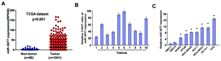
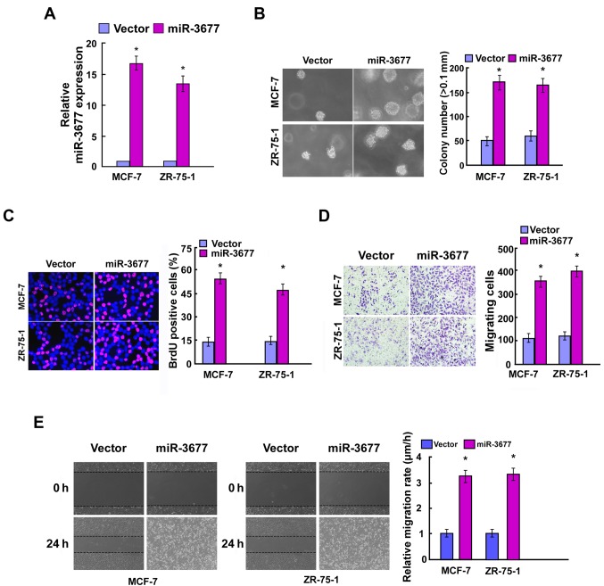
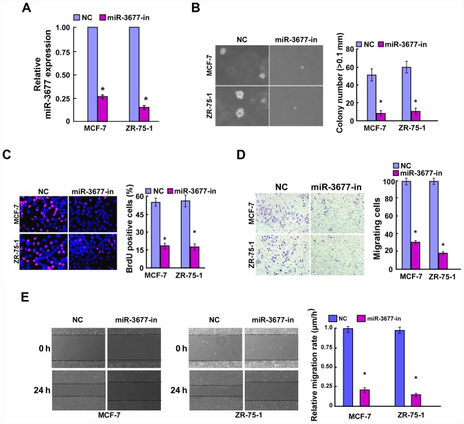
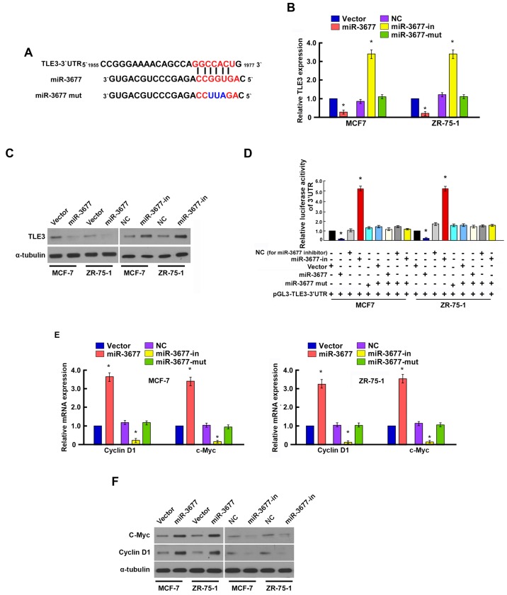
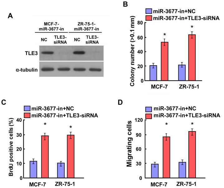
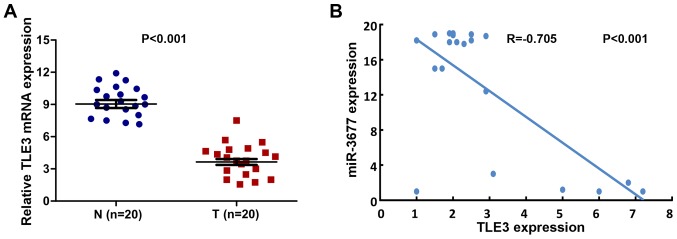
Numerous studies have indicated an important function of microRNAs (miRs) in breast cancer (BC) progression, oncogenesis and metastasis. However, the function of miR-3677, which has been revealed to be upregulated in BC [The Cancer Genome Atlas (TCGA) data], has not been investigated to date. In the present study, miR-3677 was revealed to be upregulated in BC as determined using TCGA. miR-3677 was significantly upregulated in BC tissues and cell lines compared with those noted in adjacent non-cancerous tissues and primary normal breast cells (P<0.05). The overexpression of miR-3677 promoted the cell proliferation, migration and invasion of BC cells. Using bioinformatics algorithms and luciferase assays, a novel target gene for miR-3677, namely transducin-like enhancer of Split3 (TLE3), was identified. Silencing of TLE3 in miR-3677-transfected BC cells suppressed their proliferation and migration. An inverse correlation was observed between miR-3677 and TLE3 expression levels in human BC tissues. In conclusion, the present study demonstrated that miR-3677 promoted BC cell proliferation, migration and invasion by inhibiting TLE3 expression, which provided a novel mechanism and a promising therapeutic target for patients with BC.

Keywords: breast cancer; cell metastasis; cell proliferation; microRNA-3677; transducin-like enhancer of Split3.


Figures

Figure 1.
Figure 1.
Expression of miR-3677 in human BC tissues and cell lines. (A) Expression levels of miR-3677 in BC tissues from TCGA dataset (P<0.001). (B) Relative miR-3677 expression levels in 10 paired primary BC tissues and the tumor adjacent normal tissues from the same patient were detected by RT-qPCR analysis. (C) RT-qPCR analysis of miR-3677 expression in NBECs and BC cell lines, including SKBR3, BT549, MDA-MB453, MCF-7, MDA-MB231, ZR-75-1 and T47D. Each bar represents the mean of three independent experiments. *P<0.05 vs. NBECs. miR, microRNA; BC, breast cancer; TCGA, The Cancer Genome Atlas; T, tumor tissues; ANT, adjacent normal tissues; RT-qPCR, reverse transcription-quantitative polymerase chain reaction; NBEC, normal breast cells.
Figure 2.
Figure 2.
miR-3677 upregulation promoted the cell proliferation, migration and invasion of BC. (A) Quantification of miR-3677 expression levels following transfection by polymerase chain reaction analysis. (B) Representative micrographs (left) and quantification (right) of crystal violet-stained cell colonies. (C) Representative micrographs (left) and quantification (right) of the BrdU incorporation assay in MCF-7 and ZR-75-1 cells. (D) Effect of miR-3677 overexpression in MCF-7 and ZR-75-1 cells on cell migration was analyzed using Transwell migration analyses. (E) Representative images of the wound healing assay conducted with MCF-7 and ZR-75-1 cells. Each bar represents the mean of three independent experiments. *P<0.05 vs. vector. miR, microRNA; BC, breast cancer; BrdU, bromodeoxyuridine.
Figure 3.
Figure 3.
Inhibition of miR-3677 inhibited cell proliferation, migration and invasion of BC. (A) Validation of miR-3677 expression levels following transfection by a polymerase chain reaction analysis. (B) Representative micrographs (left) and quantification (right) of crystal violet-stained cell colonies. (C) Representative micrographs (left) and quantification (right) of the BrdU incorporation assay in MCF-7 and ZR-75-1 cells. (D) Effects of miR-3677 overexpression in MCF-7 and ZR-75-1 cells on cell migration were analyzed by Transwell migration analyses. (E) Representative images of the wound healing assay conducted with MCF-7 and ZR-75-1 cells. Each bar represents the mean of three independent experiments. *P<0.05 vs. NC. miR, microRNA; BC, breast cancer; NC, negative control; BrdU, bromodeoxyuridine; in, inhibitor.
Figure 4.
Figure 4.
MiR-3677 suppresses TLE3 expression by directly targeting the TLE3 3′-UTR and altered levels of associated proteins in MCF-7 and ZR-75-1 cells. (A) Predicted miR-3677 target sequence in the 3′-UTR of TLE3 and positions of three mutated nucleotides (red) in the 3′-UTR of miR-3677 (miR-3677 mut). (B) TLE3 mRNA expression in MCF-7 and ZR-75-1 cells transfected with miR-3677 or miR-3677-in or miR-3677-mut were detected using RT-qPCR analysis. (C) Luciferase reporter assay of MCF-7 and ZR-75-1 cells transfected with the pGL3-TLE3-3′-UTR reporter and miR-3677 or miR-3677-in or miR-3677-mut or NC. (D) TLE3 protein expression in MCF-7 and ZR-75-1 cells transfected with miR-3677 or miR-3677-in or miR-3677-mut were detected by western blotting analysis. α-tubulin served as the loading control. (E) RT-qPCR analysis of expression of cyclin D1 and c-myc in MCF-7 and ZR-75-1 cells. (F) Cyclin D1 and c-Myc were measured by western blotting in MCF-7 and ZR-75-1 cells. α-tubulin served as the loading control. *P<0.05 miR-3677 vs. vector or miR-3677-in vs. NC. miR, microRNA; TLE3, transducin-like enhancer of Split3; UTR, untranslated region; RT-qPCR, reverse transcription-quantitative polymerase chain reaction; NC, negative control; in, inhibitor.
Figure 5.
Figure 5.
Downregulation of TLE3 counteracted the proliferation and cell migration inhibition caused by miR-3677-in in BC cells. (A) Western blotting verified that silencing TLE3 effectively decreased the expression of TLE3 in miR-3677-in-transfected MCF-7 and ZR-75-1 cells. (B) MiR-3677-in-transfected MCF-7 and ZR-75-1 cells following transfection with TLE3-siRNAs had increased cell colony formation. (C) BrdU proliferation assay revealed that miR-3677-in-transfected MCF-7 and ZR-75-1 cells following transfection with TLE3-siRNAs had significantly increased BrdU-positive cells. (D) MiR-3677-in-transfected MCF-7 and ZR-75-1 cells following transfection with TLE3-siRNAs had increased cell migration. Representative quantification of colonies that were >0.1 mm. Each bar represents the mean of three independent experiments. *P<0.05 vs. miR-3677-in+NC. TLE3, transducin-like enhancer of Split3; BC, breast cancer; siRNA, small interfering-RNA; BrdU, bromodeoxyuridine; NC, negative control; in, inhibitor.
Figure 6.
Figure 6.
MiR-3677 and TLE3 are inversely correlated in human breast cancer tissues. (A) Mean expression level of TLE3 in human breast cancer specimens compared with non-cancer biopsy samples. (B) In the mRNA levels, a significant inverse correlation was observed following the correlation of TLE3 with miR-3677 expression levels in the 20 human breast cancer specimens (Spearman's correlation analysis, r=−0.705; P<0.001).

References

    1. Peairs KS, Choi Y, Stewart RW, Sateia HF. Screening for breast cancer. Semin Oncol. 2017;44:60–72. doi: 10.1053/j.seminoncol.2017.02.004. - DOI - PubMed
    1. Miller KD, Siegel RL, Lin CC, Mariotto AB, Kramer JL, Rowland JH, Stein KD, Alteri R, Jemal A. Cancer treatment and survivorship statistics, 2016. CA Cancer J Clin. 2016;66:271–289. doi: 10.3322/caac.21349. - DOI - PubMed
    1. Reddy KB. MicroRNA (miRNA) in cancer. Cancer Cell Int. 2015;15:38. doi: 10.1186/s12935-015-0185-1. - DOI - PMC - PubMed
    1. Areeb Z, Stylli SS, Koldej R, Ritchie DS, Siegal T, Morokoff AP, Kaye AH, Luwor RB. MicroRNA as potential biomarkers in glioblastoma. J Neurooncol. 2015;125:237–248. doi: 10.1007/s11060-015-1912-0. - DOI - PubMed
    1. Ohtsuka M, Ling H, Doki Y, Mori M, Calin GA. MicroRNA processing and human cancer. J Clin Med. 2015;4:1651–1667. doi: 10.3390/jcm4081651. - DOI - PMC - PubMed

Publication types