. 2020 Jan 17;14(1):014108.
doi: 10.1063/1.5128739. eCollection 2020 Jan.

Chemotropism among populations of yeast cells with spatiotemporal resolution in a biofabricated microfluidic platform

Affiliations

Chemotropism among populations of yeast cells with spatiotemporal resolution in a biofabricated microfluidic platform

Thanh Vo et al. Biomicrofluidics. .

Abstract

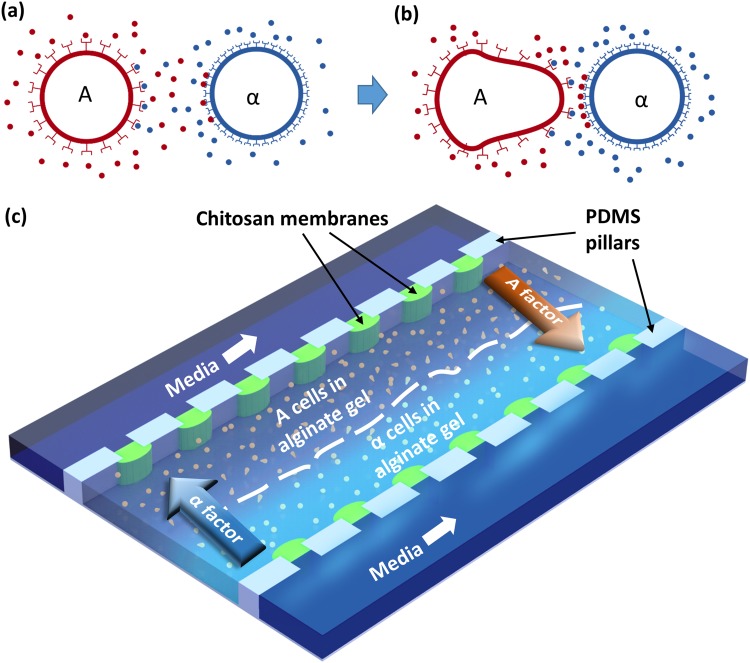
Chemotropism is an essential response of organisms to external chemical gradients that direct the growth of cells toward the gradient source. Chemotropic responses between single cells have been studied using in vitro gradients of synthetically derived signaling molecules and helped to develop a better understanding of chemotropism in multiple organisms. However, dynamic changes including spatial changes to the gradient as well as fluctuations in levels of cell generated signaling molecules can result in the redirection of chemotropic responses, which can be difficult to model with synthetic peptides and single cells. An experimental system that brings together populations of cells to monitor the population-scale chemotropic responses yet retain single cell spatiotemporal resolution would be useful to further inform on models of chemotropism. Here, we describe a microfluidic platform that can measure the chemotropic response between populations of mating yeast A- and α-cells with spatiotemporal programmability and sensitivity by positioning cell populations side by side in calcium alginate hydrogels along semipermeable membranes with micrometer spatial control. The mating phenotypes of the yeast populations were clearly observed over hours. Three distinct responses were observed depending on the distance between the A- and α-cell populations: the cells either continued to divide, arrest, and develop a stereotypical polarized projection termed a "shmoo" toward the cells of opposite mating type or formed shmoos in random directions. The results from our studies of yeast mating suggest that the biofabricated microfluidic platform can be adopted to study population-scale, spatial-sensitive cell-cell signaling behaviors that would be challenging using conventional approaches.


Figures

Scheme 1.
Scheme 1.
Mating yeast chemotropism with biofabrication in microfluidics. (a) Signaling between mating yeast A- and α-cells; (b) chemotropic growth of A-cell toward α-cell resulting in so-called “shmoo” structures, in which A-cells were deleted for BAR1, a gene that encodes for a protease that degrades α-factor to increase their sensitivity to α-cells; and (c) A- and α-cells trapped side by side in alginate hydrogels in a middle microchannel. Biofabricated semi-permeable chitosan membranes separate the middle microchannel from two-side microchannels where media solution is introduced to culture the cells.
FIG. 1.
FIG. 1.
Positioning yeast cells with biofabrication in microfluidics. (a) A PDMS microfluidic device with multiple sets of microchannel networks, with the zoom-in dash rectangle showing in (b) the biofabricated freestanding and semipermeable chitosan membranes connecting three microchannels; (c) schematic and (d) micrograph of the assembly of yeast cells in the alginate hydrogel; (e) schematic and (f) micrograph of assembled cells after PBS rinsing; (g) cells uniformly distributed in the alginate hydrogel; and (h) cells aggregated in the solution after 10 min without cross-linking the alginate solution into the hydrogel with calcium ions.
FIG. 2.
FIG. 2.
The fate of chemotropism or proliferation of mating yeast cells depended on the distance from the other cell type. (a) Schematic of the A- and α-cells assembled side by side in the alginate hydrogel in the middle channel of the three-channel device. A medium was introduced in the side microchannels to culture the cells over time; (b) relative uniform distribution of A- and α-cells at 1 h after assembly, with the area of A-cells, was separated into four blocks for further analysis in (d) and (e); (c) distribution and morphologies of A- and α-cells at 8 h after assembly, showing chemotropism of A-cells closer to α-cells while continuous proliferation of A-cells away from α-cells; (d) proliferation ratio of A-cells in the four indicated blocks in (b) and (c), showing continuous cell proliferation over 5 h in blocks 1 and 2, while little cell proliferation in block 4. Proliferation over 5 h was not accountable due to cell overlapping; and (e) Projection ratio of A-cells in the same four blocks, showing little or no chemotropism over 8 h for cells in blocks 1 and 2 while increasing chemotropism for cells in block 4.
FIG. 3.
FIG. 3.
Proliferation ratios of A-cells with chemotropism (blue rectangles and black triangles) vs those of α-cells without chemotropism (red diamonds and green circles) as the control experiment. The proliferation of A-cells was spatially sensitive depending on the distance of concerned cells from the mating opposite α-cells, while the proliferation of α-cells was spatially insensitive. The dashed lines are 2nd order polynomial fits. The data points were the ratio of exact cell counts without statistics.
FIG. 4.
FIG. 4.
Response of one representative individual A-cell vs clusters of A-cells to mating signal. (a) Time lapse images of an individual (indicated with red arrows) and clusters of A-cells near α-cells; (b) the cell proliferation ratios of clusters of A-cells compared with an individual A-cell over a time period of 4.5 h. The data points were the ratio of exact cell counts without statistics; (c) the projection ratio of an individual A-cell compared with that of clusters of A-cells over time. The error bars represent the standard deviations of three measurements of one cell (individual A-cell) or the measurements of five cells (clusters of A-cells).
FIG. 5.
FIG. 5.
Directional yeast chemotropism was pheromone gradient-dependent. (a) In the case of high density of A-cells near α-cells, the α pheromone signal was consumed by A-cells resulting in sharper gradient, thus the chemotropism of A-cells transited from non-directional (region i) to unidirectional (region ii) to little chemotropism (region iii). This is most obvious in the evolving images at 210 and 270 min. (b) In the case of low density of A-cells near α-cells, the α-factor was saturated around A-cells with little gradient, thus the chemotropism of A-cells remained non-directional.
FIG. 6.
FIG. 6.
Multi-lobed cell morphology under saturated α pheromone signal. (a) Representative multi-lobed morphology of one A-cell (red rectangle) under the configuration that a few A-cells (right) were assembled near to abundant α-cells (left); (b) Time lapse images of the representative A-cell growing into multi-lobed morphology at 3–8 h.

Similar articles

Cited by

References

    1. Cai H. and Devreotes P. N., “Moving in the right direction: How eukaryotic cells migrate along chemical gradients,” Semin. Cell Dev. Biol. 22(8), 834–841 (2011). 10.1016/j.semcdb.2011.07.020 - DOI - PMC - PubMed
    1. Tojima T., Hines J. H., Henley J. R., and Kamiguchi H., “Second messengers and membrane trafficking direct and organize growth cone steering,” Nat. Rev. Neurosci. 12(4), 191–203 (2011). 10.1038/nrn2996 - DOI - PMC - PubMed
    1. Palanivelu R. and Preuss D., “Pollen tube targeting and axon guidance: Parallels in tip growth mechanisms,” Trends Cell Biol. 10(12), 517–524 (2000). 10.1016/S0962-8924(00)01849-3 - DOI - PubMed
    1. Ismael A. and Stone D. E., “Yeast chemotropism: A paradigm shift in chemical gradient sensing,” Cell Logist. 7(2), e1314237 (2017). 10.1080/21592799.2017.1314237 - DOI - PMC - PubMed
    1. Arkowitz R. A., “Chemical gradients and chemotropism in yeast,” Cold Spring Harb. Perspect. Biol. 1(2), a001958 (2009). 10.1101/cshperspect.a001958 - DOI - PMC - PubMed

LinkOut - more resources