Review
. 2019 Dec 19;9(1):1703244.
doi: 10.1080/20013078.2019.1703244. eCollection 2020.

Inhibiting extracellular vesicles formation and release: a review of EV inhibitors

Affiliations
Review

Inhibiting extracellular vesicles formation and release: a review of EV inhibitors

Mariadelva Catalano et al. J Extracell Vesicles. .

Abstract

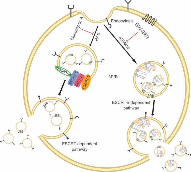
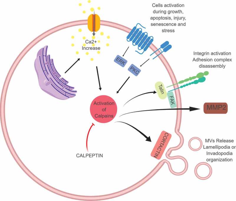
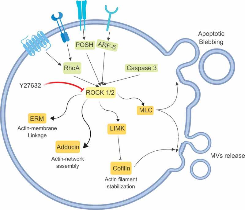
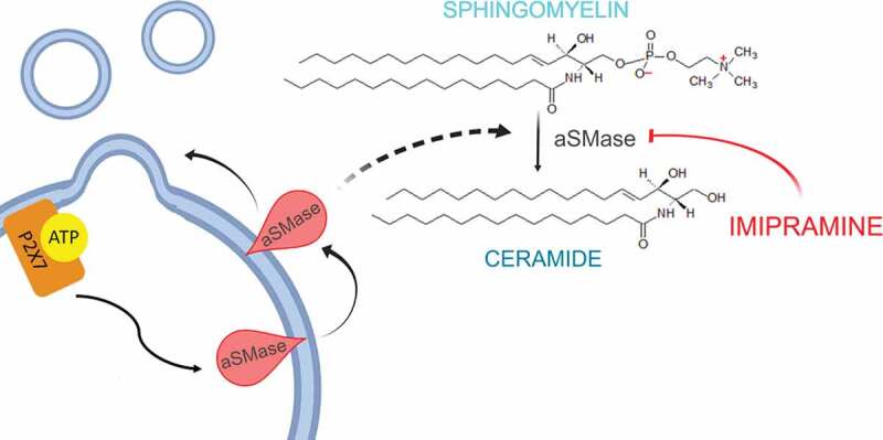
It is now becoming well established that vesicles are released from a broad range of cell types and are involved in cell-to-cell communication, both in physiological and pathological conditions. Once outside the cell, these vesicles are termed extracellular vesicles (EVs). The cellular origin (cell type), subcellular origin (through the endosomal pathway or pinched from the cell membrane) and content (what proteins, glycoproteins, lipids, nucleic acids, metabolites) are transported by the EVs, and their size, all seem to be contributing factors to their overall heterogeneity. Efforts are being invested into attempting to block the release of subpopulations of EVs or, indeed, all EVs. Some such studies are focussed on investigating EV inhibitors as research tools; others are interested in the longerterm potential of using such inhibitors in pathological conditions such as cancer. This review, intended to be of relevance to both researchers already well established in the EV field and newcomers to this field, provides an outline of the compounds that have been most extensively studied for this purpose, their proposed mechanisms of actions and the findings of these studies.

Keywords: Extracellular vesicles; GW4869; Y27632; calpeptin; exosomes; imipramine; inhibitors; manumycin A; microvesicles; pantethine.


Figures

Figure 1.
Figure 1.
Exosomes are released from intracellular compartments known as multi-vesicular bodies (MVBs). MVBs biogenesis is associated with two different mechanisms: ESCRT-dependent and ESCRT-independent pathways.
Figure 2.
Figure 2.
Calpains, once activated through calcium binding, can activate different cellular processes including cell migration (through their interaction with Talin and FAK), cell invasion (thorough the promotion of MMP release and activation), and MVs formation and release (through their activity on cytoskeletal proteins including cortactin). Calpain inhibitors, of which calpeptin is one of the most studied, are under investigation for clinical application.
Figure 3.
Figure 3.
Rho-associated protein kinases (ROCK) are serine-threonine kinases involved in cytoskeleton re-organisation. Once activated by multiple stimuli, ROCK regulate the shape and the movement of the cells, through the activation of Adducin or ERM (ezrin, radixin and moesin), but they can also interact with MLC (myosin light chain) and LIMK (LIM kinases, able to inactivate cofilin, fundamental for actin filament stabilisation) both involved in MVs release. Y27632 is a competitive inhibitor of both ROCK1 and ROCK2 and by blocking these proteins it can inhibit MVs release.
Figure 4.
Figure 4.
MVs present unique lipid characteristics, including an enrichment of sphingomyelin and ceramide, thus every enzyme able to interfere with membrane composition, i.e. calpains, scramblases and acid sphingomyelinases, plays a key role in MVs biogenesis. Acid sphingomyelinases convert sphingomyelin into ceramide, a cone-shaped rigid lipid that forms micro-domains inside the cell membrane, inducing the budding of MVs. Imipramine, a well-known anti-depressant, can promote membrane fluidity by acting on aSMases, thus preventing MVs generation.

Similar articles

Cited by

References

    1. Yuana Y, Sturk A, Nieuwland R. Extracellular vesicles in physiological and pathological conditions. Blood Rev. 2013:27:31–39. doi:10.1016/j.blre.2012.12.002 - DOI - PubMed
    1. van der Pol E, Böing AN, Harrison P, et al. Classification, functions, and clinical relevance of extracellular vesicles. Pharmacol Rev. 2012;64:676–22. - PubMed
    1. Minciacchi VR, Freeman MR, Di Vizio D.. Extracellular vesicles in cancer: exosomes, microvesicles and the emerging role of large oncosomes. Semin Cell Dev Biol. 2015;40:41–51. - PMC - PubMed
    1. Huotari J, Helenius A.. Endosome maturation. Embo J. 2011;30:3481–3500. - PMC - PubMed
    1. Chiaruttini N, Redondo-Morata L, Colom A, et al. Relaxation of loaded ESCRT-III spiral springs drives membrane deformation. Cell. 2015;163:866–879. - PMC - PubMed

LinkOut - more resources