

Phenotypic analysis of extracellular vesicles: a review on the applications of fluorescence
- PMID: 32002172
- PMCID: PMC6968689
- DOI: 10.1080/20013078.2019.1710020
Phenotypic analysis of extracellular vesicles: a review on the applications of fluorescence
Abstract
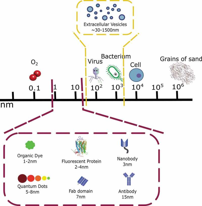
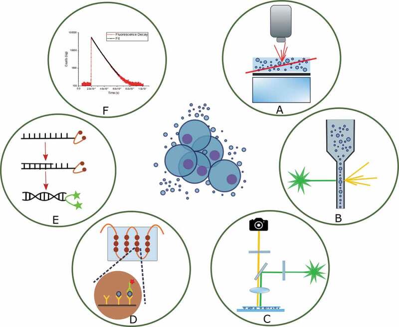
Extracellular vesicles (EVs) have numerous potential applications in the field of healthcare and diagnostics, and research into their biological functions is rapidly increasing. Mainly because of their small size and heterogeneity, there are significant challenges associated with their analysis and despite overt evidence of the potential of EVs in clinical diagnostic practice, guidelines for analytical procedures have not yet been properly established. Here, we present an overview of the main methods for studying the properties of EVs based on the principles of fluorescence. Setting aside the isolation, purification and physicochemical characterization strategies which answer questions about the size, surface charge and stability of EVs (reviewed elsewhere), we focus on available optical tools that enable the direct analysis of phenotype and mechanisms of interaction with tissues. In brief, the topics on which we elaborate range from the most popular approaches such as nanoparticle tracking analysis and flow cytometry, to less commonly used techniques such as fluorescence depolarization and microarrays as well as emerging areas such as fast fluorescence lifetime imaging microscopy (FLIM). We highlight that understanding the strengths and limitations of each method is essential for choosing the most appropriate combination of analytical tools. Finally, future directions of this rapidly developing area of medical diagnostics are discussed.
Keywords: Exosomes; FLIM; FRET; cancer; fluorescence depolarization; fluorescence lifetime; fluorescent dye; fluorescent protein; microarrays; microfluidics; microscopy; nanobody; nanoparticle tracking analysis; qPCR; quantum dot; super resolution microscopy.
© 2020 The Author(s). Published by Informa UK Limited, trading as Taylor & Francis Group.
Figures
Similar articles
-
Enumeration of extracellular vesicles by a new improved flow cytometric method is comparable to fluorescence mode nanoparticle tracking analysis.Nanomedicine. 2016 May;12(4):977-986. doi: 10.1016/j.nano.2015.12.370. Epub 2016 Jan 6. Nanomedicine. 2016. PMID: 26767510
-
An Emerging Fluorescence-Based Technique for Quantification and Protein Profiling of Extracellular Vesicles.SLAS Technol. 2021 Apr;26(2):189-199. doi: 10.1177/2472630320970458. Epub 2020 Nov 13. SLAS Technol. 2021. PMID: 33185120
-
Fluorescent labeling of extracellular vesicles.Methods Enzymol. 2020;645:15-42. doi: 10.1016/bs.mie.2020.09.002. Epub 2020 Sep 29. Methods Enzymol. 2020. PMID: 33565969
-
Microfluidic approaches for isolation, detection, and characterization of extracellular vesicles: Current status and future directions.Biosens Bioelectron. 2017 May 15;91:588-605. doi: 10.1016/j.bios.2016.12.062. Epub 2016 Dec 30. Biosens Bioelectron. 2017. PMID: 28088752 Free PMC article. Review.
-
Recent advances on protein-based quantification of extracellular vesicles.Anal Biochem. 2021 Jun 1;622:114168. doi: 10.1016/j.ab.2021.114168. Epub 2021 Mar 16. Anal Biochem. 2021. PMID: 33741309 Review.
Cited by
-
Rapid Characterization and Quantification of Extracellular Vesicles by Fluorescence-Based Microfluidic Diffusion Sizing.Adv Healthc Mater. 2022 Mar;11(5):e2100021. doi: 10.1002/adhm.202100021. Epub 2021 Jun 10. Adv Healthc Mater. 2022. PMID: 34109753 Free PMC article.
-
Unravelling Plasma Extracellular Vesicle Diversity With Optimised Spectral Flow Cytometry.J Extracell Biol. 2025 Apr 25;4(4):e70045. doi: 10.1002/jex2.70045. eCollection 2025 Apr. J Extracell Biol. 2025. PMID: 40292386 Free PMC article.
-
Emerging Biomimetic Drug Delivery Nanoparticles Inspired by Extracellular Vesicles.Wiley Interdiscip Rev Nanomed Nanobiotechnol. 2025 Jul-Aug;17(4):e70025. doi: 10.1002/wnan.70025. Wiley Interdiscip Rev Nanomed Nanobiotechnol. 2025. PMID: 40840553 Free PMC article. Review.
-
A Microfluidic Platform to Monitor Real-Time Effects of Extracellular Vesicle Exchange between Co-Cultured Cells across Selectively Permeable Barriers.Int J Mol Sci. 2022 Mar 24;23(7):3534. doi: 10.3390/ijms23073534. Int J Mol Sci. 2022. PMID: 35408896 Free PMC article.
-
Mesenchymal Stromal Cell-Derived Extracellular Vesicles as Biological Carriers for Drug Delivery in Cancer Therapy.Front Bioeng Biotechnol. 2022 Apr 14;10:882545. doi: 10.3389/fbioe.2022.882545. eCollection 2022. Front Bioeng Biotechnol. 2022. PMID: 35497332 Free PMC article. Review.
References
-
- Wolf P. The nature and significance of platelet products in human plasma. Br J Haematol. 1967;13:269–18. - PubMed
-
- Nieuwland R, Sturk A.. Why do cells release vesicles? Thromb Res. 2010;125:S49–S51. - PubMed
-
- Théry C, Zitvogel L, Amigorena S. Exosomes: composition, biogenesis and function. Nat Rev Immunol. 2002;2:569. - PubMed
-
- Colombo M, Raposo G, Théry C. Biogenesis, secretion, and intercellular interactions of exosomes and other extracellular vesicles. Annu Rev Cell Dev Biol. 2014;30:255–289. - PubMed
Publication types
LinkOut - more resources
Full Text Sources
