. 2020 Jan 7;9(1):1710899.
doi: 10.1080/20013078.2019.1710899. eCollection 2020.

Tracking the evolution of circulating exosomal-PD-L1 to monitor melanoma patients

Affiliations

Tracking the evolution of circulating exosomal-PD-L1 to monitor melanoma patients

Marine Cordonnier et al. J Extracell Vesicles. .

Abstract

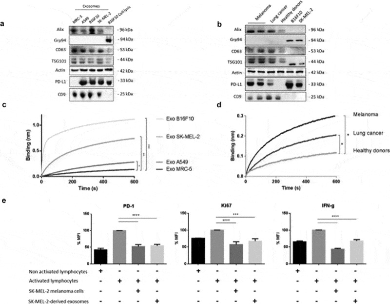
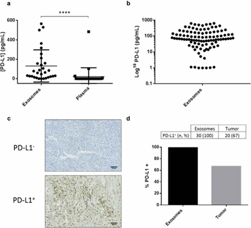
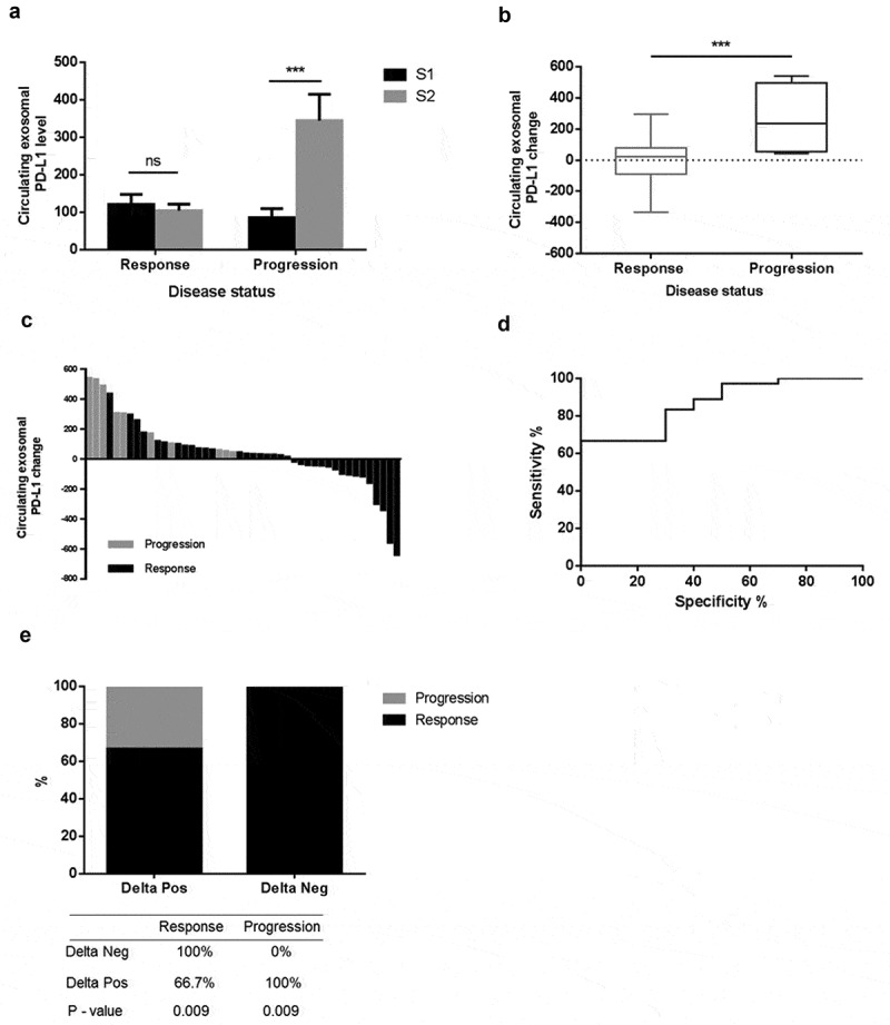
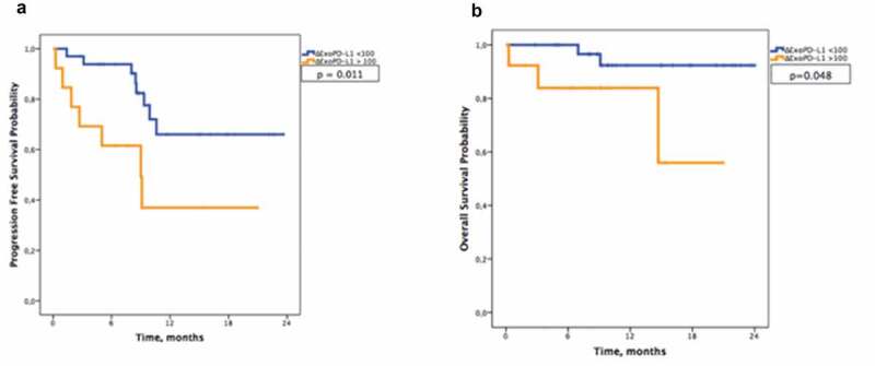
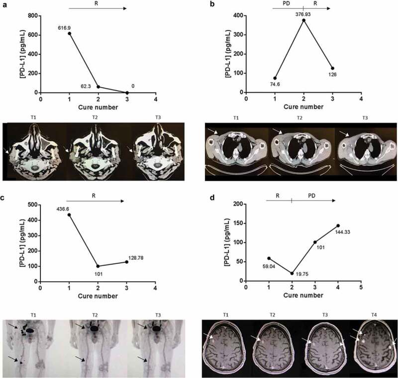
In the era of immunotherapies there is an urgent need to implement the use of circulating biomarkers in clinical practice to facilitate personalized therapy and to predict treatment response. We conducted a prospective study to evaluate the usefulness of circulating exosomal-PD-L1 in melanoma patients' follow-up. We studied the dynamics of exosomal-PD-L1 from 100 melanoma patients by using an enzyme-linked immunosorbent assay. We found that PD-L1 was secreted through exosomes by melanoma cells. Exosomes carrying PD-L1 had immunosuppressive properties since they were as efficient as the cancer cell from which they derive at inhibiting T-cell activation. In plasma from melanoma patients, the level of PD-L1 (n= 30, median 64.26 pg/mL) was significantly higher in exosomes compared to soluble PD-L1 (n= 30, 0.1 pg/mL). Furthermore, exosomal-PD-L1 was detected in all patients whereas only 67% of tumour biopsies were PD-L1 positive. Although baseline exosomal-PD-L1 levels were not associated with clinic-pathologic characteristics, their variations after the cures (ΔExoPD-L1) correlated with the tumour response to treatment. A ΔExoPD-L1 cut-off of> 100 was defined, yielding an 83% sensitivity, a 70% specificity, a 91% positive predictive value and 54% negative predictive values for disease progression. The use of the cut-off allowed stratification in two groups of patients statistically different concerning overall survival and progression-free survival. PD-L1 levels in circulating exosomes seem to be a more reliable marker than PD-L1 expression in tumour biopsies. Monitoring of circulating exosomal-PD-L1 may be useful to predict the tumour response to treatment and clinical outcome.

Keywords: Melanoma; PD-L1/PD-1; exosome; follow-up; immune checkpoint.


Conflict of interest statement

No potential conflict of interest was reported by the authors.

Figures

Figure 1.
Figure 1.
PD-L1 expression in tumour-derived exosomes. (A/B) Representative immunoblots showing expression of Alix, CD63, TSG101, PD-L1 and CD9 in exosomes derived from cell lines (a) MRC-5 (normal human lung cells), A549 (human lung cancer), B16F10 (mouse melanoma) or SK-MEL-2 (human melanoma) or (b) plasma-derived exosomes from melanoma patients, lung cancer patients and healthy donors. Grp94 is used here as an exosomal negative control and actin as a loading control. Cell lysates are also included. (C/D) Binding of exosomes (nm) derived from cell lines (MRC-5, A549, SK-MEL-2, B16F10) or from plasma samples of melanoma patients (n = 6) or lung cancer patients (n = 6) and healthy donors (n = 5) to immobilized biotinylated PD-1 determined by biolayer interferometry. Binding curves represent mean signal of triplicate measurements for each sample. Mann–Whitney. (c), ***p < 0.001, **p = 0.0081, *p = 0.0041. (d) *p = 0.0039 melanoma, *p = 0.0031 lung cancer). (e) Percentage of PD-1, Ki67 and IFNϒ mean fluorescence intensity in lymphocytes cultured 24 h in the presence or absence of SK-MEL-2 or SK-MEL-2-derived exosomes, determined by flow cytometry (***p = 0.0006, ****p <0.0001).
Figure 2.
Figure 2.
PD-L1 is easily detected in exosomes, when compared with soluble PD-L1 in plasma or in tumour biopsies. (a) Levels of PD-L1 in exosomes isolated from the plasma of melanoma patients compared with PD-L1 levels free in the plasma (n = 30) (****p < 0.0001), determined by ELISA. (b) Levels of ExoPD-L1 isolated from the 100 patient plasma samples of the EXOMEL cohort. (c) Representative IHC image of PD-L1 negative (PD-L1) or positive (PD-L1+) tumours (22C3 antibody). Scale bars indicated 100 µm. (d) Percentage of patients positive for PD-L1 when measured in circulating exosomes versus tumour biopsies.
Figure 3.
Figure 3.
Changes in the level of ExoPD-L1 stratify melanoma patients according to disease status. (a) Mean value of circulating ExoPD-L1 evaluated at S1 and S2 in patients grouped according to disease status (response, n = 36) or progression, n = 10). Mann–Whitney (***p = 0.0002). (b) Comparison of changes in level of ExoPD-L1 in melanoma patients between S1/S2, according to disease status. Mann–Whitney (***p = 0.0002). (c) Waterfall plots showing changes in level of ExoPD-L1 between S1/S2, according to disease status. Black bars represent responder patients (n = 36) and grey bars represent progressive patients (n = 10). (d) ROC curve analysis of changes in level of ExoPD-L1 (S1/S2) in responder patients compared with non-responders (AUC = 0.867, SE 0.057, Cl95% 0.755–0.978; p < 0.001). (e) An increase in ExoPD-L1 is associated with disease progression p < 0.009. A decrease in ExoPD-L1 is associated with response p < 0.009. All patients with a decrease in level of ExoPD-L1 experienced a tumour response and all patients with an increase in level of ExoPD-L1 experienced progression.
Figure 4.
Figure 4.
Melanoma-derived ExoPD-L1 can be used as a marker of survival. Kaplan–Meier estimates of (a) progression-free survival (p = 0.011) and (b) overall survival (p = 0.048) in patients according to ΔExoPD-L1 (n = 46).
Figure 5.
Figure 5.
ExoPD-L1 can be used as follow-up markers in melanoma patients. Case study of the correlation between ExoPD-L1 levels in plasma samples from melanoma patients and response to PD-L1-based therapy. Concomitant imaging and ExoPD-L1 sampling in four patients experiencing (a) response as observed in the parotid gland and cervical lymph nodes on CT scan; (b) complete response in the subcutaneous tissue, popliteal and ilioinguinal lymph nodes on PET-CT; (c) disease response in the muscle and infraclavicular lymph nodes on CT scan; (d) initial response (at cure 1) then disease progression as detected in the brain on MRI. R: response, PD: progression of the disease. Tumour metastases in the scans are indicated by an arrow.

References

    1. Topalian SL, Hodi FS, Brahmer JR, et al. Safety, activity, and immune correlates of anti-pd-1 antibody in cancer. N Engl J Med. 2012;366(26):2443–2411. - PMC - PubMed
    1. Ribas A, Hamid O, Daud A, et al. Association of pembrolizumab with tumor response and survival among patients with advanced melanoma. JAMA. 2016. April 19;315(15):1600–1609. - PubMed
    1. Robert C, Long GV, Brady B, et al. Nivolumab in previously untreated melanoma without BRAF mutation. N Engl J Med. 2015. January 22;372(4):320–330. - PubMed
    1. Kaunitz GJ, Cottrell TR, Lilo M, et al. Melanoma subtypes demonstrate distinct PD-L1 expression profiles. Lab Investig J Tech Methods Pathol. 2017. September;97(9):1063–1071. - PMC - PubMed
    1. Madore J, Vilain RE, Menzies AM, et al. PD-L1 expression in melanoma shows marked heterogeneity within and between patients: implications for anti-PD-1/PD-L1 clinical trials. Pigment Cell Melanoma Res. 2015. May;28(3):245–253. - PubMed

LinkOut - more resources