

doi: 10.1093/af/vfz024.
eCollection 2019 Jul.
The development of brain magnetic resonance approaches in large animal models for preclinical research
Affiliations
- PMID: 32002261
- PMCID: PMC6951960
- DOI: 10.1093/af/vfz024
Item in Clipboard
The development of brain magnetic resonance approaches in large animal models for preclinical research
Anim Front.
.
No abstract available
Keywords: brain; image analysis; magnetic resonance imaging; pathology.
Figures
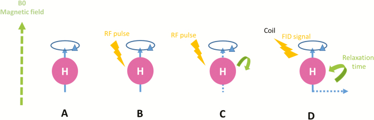
Schematic representation of magnetic resonance imaging principles. (A) When placed in a strong magnetic field B0, protons align themselves parallel or antiparallel with the direction of B0. (B) The application of a RF pulse B1 perpendicular to B0 and at the same frequency of B0 allows the longitudinal magnetization to tilt away from B0 (resonance). (C) Once the RF signal stops, M returns to equilibrium (relaxation) such that it aligns again with B0. (D) During relaxation, protons lose energy by emitting a RF signal Free Induction Decay (FID) which can be measured by a coil placed around the object being imaged and subsequently reconstructed to obtain 3D gray-scale MR images.
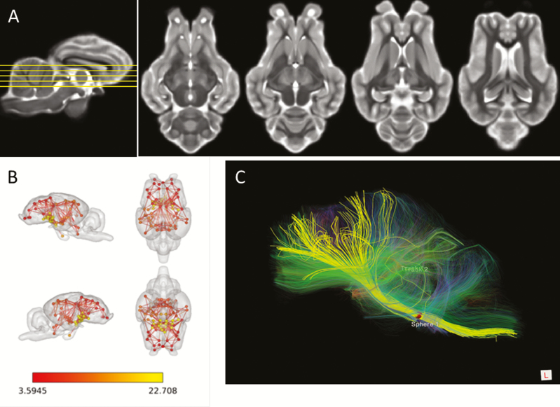
The multimodal potency of MRI. (A) T2 weighed MRI images of the sheep brain at four different dorso–ventral levels. (B) Resting-state networks defined using resting state fMRI and associated analysis in anesthetized sheep. Three internal subnetworks involving the frontal region (red nodes), the occipito–parietal region (orange nodes), and the diencephalic region (yellow nodes) can be defined. (C) Identification of some main fiber tracts (yellow) connecting the brainstem (right) to the frontal regions (left) and visualized by diffusion tensor imaging within the sheep brain.
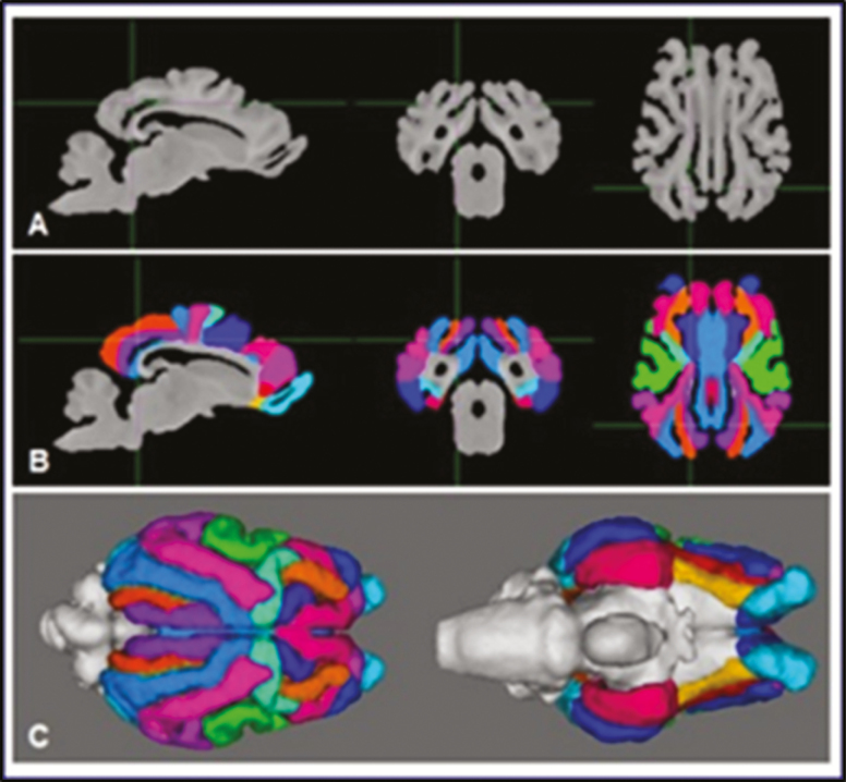
Steps for the realization of a sheep brain template and atlas. (A) Acquisition of brain MR images and computation of a MR template of the sheep brain in T1-weighted contrast. (B) Segmentation of cortical areas overlaid on the template images. (C) Three-dimensional view of the segmented telencephalon.
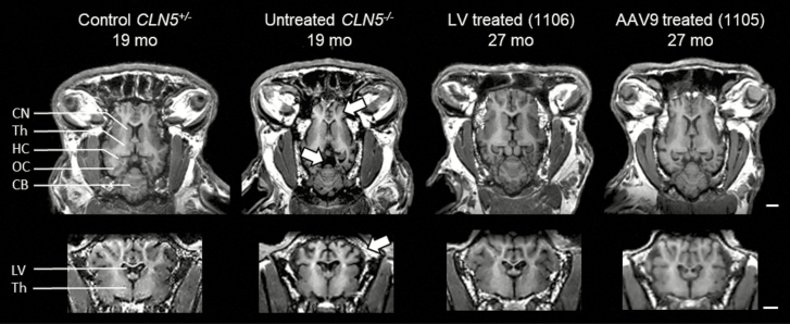
T1-weighted MRI images showing the preservation of neuroanatomical structures after gene therapy in CLN5−/− sheep. T1-weighted MRI images in the horizontal (top) or coronal (bottom) views from 19-mo old control or affected sheep compared with much older (27 mo old) sheep treated by gene therapy with lentivirus (LV) or single-stranded adeno-associated viral serotype 9 (AAV9) before the development of clinical signs. The treatments protected from the high neuronal atrophy observed in affected animals because either vector preserved neuroanatomical structures, protecting against the profound cortical atrophy (top arrow), prominent ventricular enlargement (center arrow), and cranial thickening (bottom arrow) seen in the untreated CLN5−/− animals. CB = cerebellum; CN = caudate nucleus; HC = hippocampus; LV = lateral ventricles; OC = occipital cortex; Th = thalamus. By permission from Elsevier (original publication: Mitchell et al. (2018), Molecular Therapy, 26(10): 2366–2378).
References
-
- Baker E.W., Platt S.R., Lau V.W., Grace H.E., Holmes S.P., Wang L., Duberstein K.J., Howerth E.W., Kinder H.A., Stice S.L., . et al. 2017. Induced pluripotent stem cell-derived neural stem cell therapy enhances recovery in an ischemic stroke pig model. Sci. Rep. 7:10075. doi: 10.1038/s41598-017-10406-x - DOI - PMC - PubMed
-
- Boltze J., Ferrara F., Hainsworth A.H., Bridges L.R., Zille M., Lobsien D., Barthel H., Mc Leod D.D., Grässer F., Pietsch S., . et al. 2018. Lesional and perilesional tissue characterization by automated image processing in a novel gyrencephalic animal model of peracute intracerebral hemorrhage. J. Cereb. Blood Flow Metab., in press. doi: 10.1177/027/1678X18802119 - DOI - PMC - PubMed
LinkOut - more resources
Full Text Sources