

Multimodal Imaging and Histopathological Evaluation of Berger's Space
- PMID: 32002397
- PMCID: PMC6984151
- DOI: 10.1159/000495724
Multimodal Imaging and Histopathological Evaluation of Berger's Space
Abstract
Objective: To demonstrate the multimodal imaging and histopathology of Berger's space.
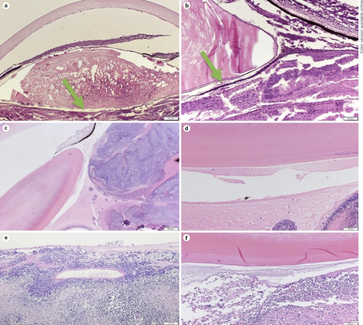
Methods: We conducted a retrospective in vivo analysis of 4 patients demonstrating Berger's space with intraocular pathological conditions, documented by slit-lamp biomicroscopic photography and, in 2 patients, also by optical coherence tomography (OCT). Additionally, we carried out a retrospective histological study of 7 enucleated eyes with retinoblastoma demonstrating Berger's space. A review of the literature was also performed.
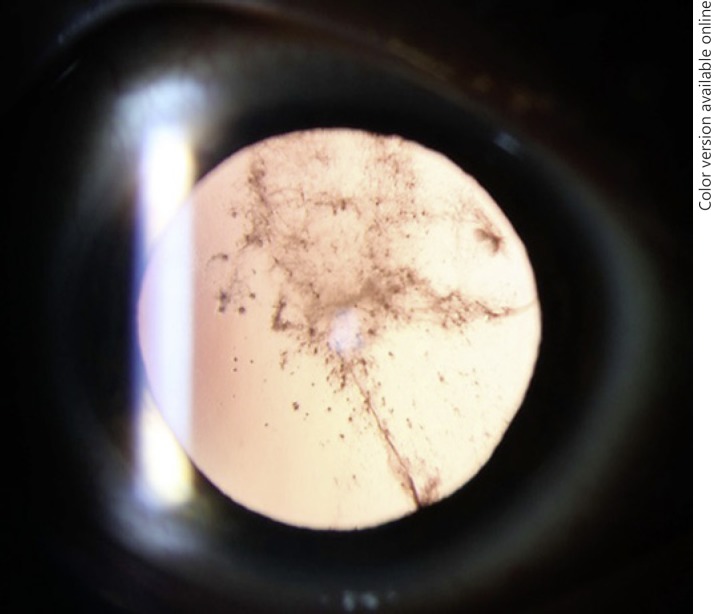
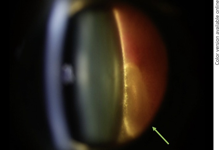
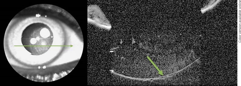
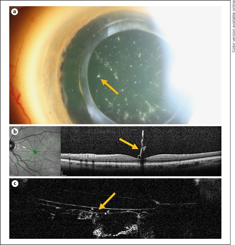
Results: Two eyes had slit-lamp photographs. One case showed Berger's space surrounded by vitreous hemorrhage. In the other case, amyloid was trapped within Berger's space. In another 2 eyes that were pseudophakic, Berger's space was visible on anterior segment OCT. One had amyloid trapped in Berger's space that could be seen with OCT. The histological review of the 7 enucleated eyes with advanced retinoblastoma demonstrated the presence of pyknotic cells in Berger's space.
Conclusions: Berger's space is an actual space in pathological conditions and can be an important site of pathology. Additionally, to our knowledge, this is the first time that Berger's space has been documented by anterior segment OCT in a clinical setting.
Keywords: Amyloid; Berger's space; Optical coherence tomography; Vitreous.
Copyright © 2019 by S. Karger AG, Basel.
Conflict of interest statement
No conflicting relationship exists for V.M., M.B.N., or D.R.S. J.S.P. has stock in LAgen Laboratories, which supplies induced pluripotent stem cell-derived retinal pigment epithelial cells for in vitro studies and has no relevant financial interests.
Figures
References
-
- Berger E. Wiesbaden: Bergmann; 1887. Beitrage zur Anatomie des Auges in Normalem und Pathologischem Zustande.
-
- Wieger G. Über den Canalis Petiti und ein ‘Ligamentum hyaloideocapsulare'; thesis. Strassurg. 1883
-
- Tassignon MJ, Ní Dhubhghaill S. Real-Time Intraoperative Optical Coherence Tomography Imaging Confirms Older Concepts About the Berger Space. Ophthalmic Res. 2016;56((4)):222–226. - PubMed
-
- Turgut B, Türkçüoğlu P, Deniz N, Catak O. Annular and central heavy pigment deposition on the posterior lens capsule in the pigment dispersion syndrome: pigment deposition on the posterior lens capsule in the pigment dispersion syndrome. Int Ophthalmol. 2008 Dec;28((6)):441–5. - PubMed
Publication types
LinkOut - more resources
Full Text Sources
