. 2020 May;30(5):2463-2472.
doi: 10.1007/s00330-019-06609-7. Epub 2020 Jan 30.

Minimal ablative margin (MAM) assessment with image fusion: an independent predictor for local tumor progression in hepatocellular carcinoma after stereotactic radiofrequency ablation

Affiliations

Minimal ablative margin (MAM) assessment with image fusion: an independent predictor for local tumor progression in hepatocellular carcinoma after stereotactic radiofrequency ablation

Gregor Laimer et al. Eur Radiol. 2020 May.

Abstract

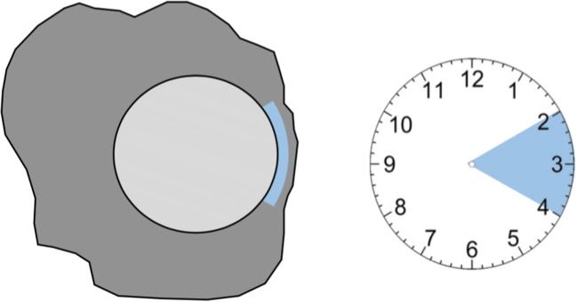
Objectives: To assess the minimal ablative margin (MAM) by image fusion of intraprocedural pre- and post-ablation contrast-enhanced CT images and to evaluate if it can predict local tumor progression (LTP) independently. Furthermore, to determine a MAM with which a stereotactic radiofrequency ablation (SRFA) can be determined successful and therefore used as an intraprocedural tool to evaluate treatment success.

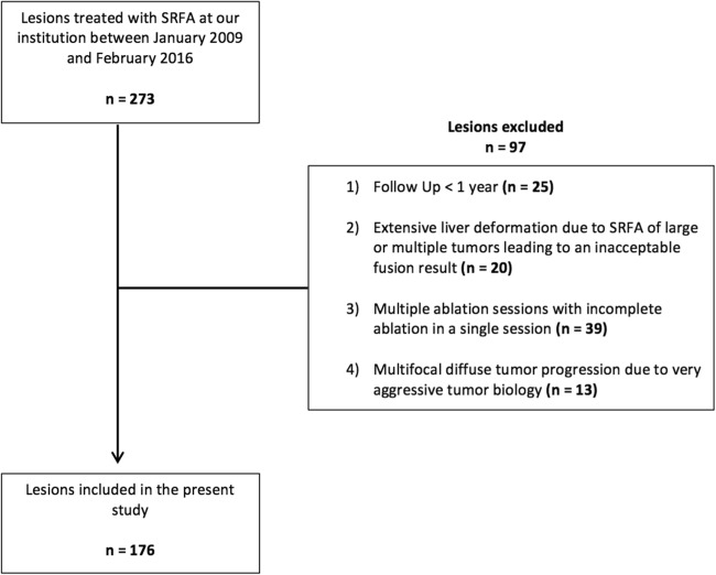
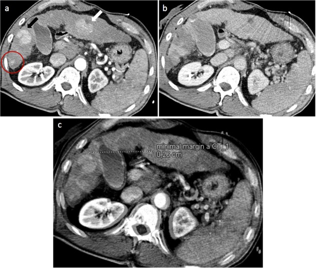
Methods: A total of 110 patients (20 women, 90 men; mean age 63.7 ± 10.2) with 176 hepatocellular carcinomas were assessed by retrospective analysis of prospectively collected data. The MAM was determined through image fusion of intraprocedural pre- and post-ablation images using commercially available rigid imaging registration software. LTP was assessed in contrast-enhanced CTs or MR scans at 3-6-month intervals.

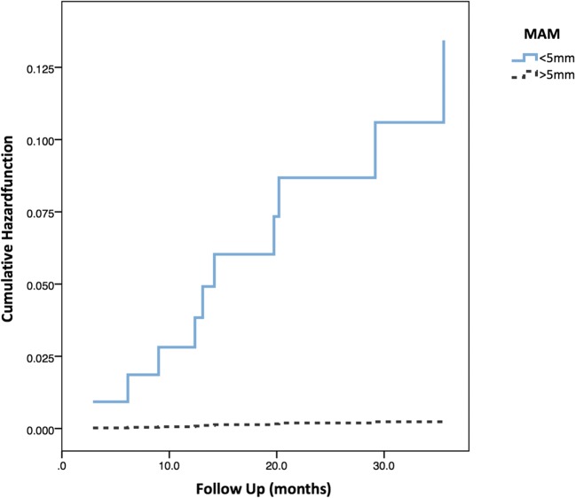
Results: The MAM was the only significant independent predictor of LTP (p = 0.036). For each millimeter increase of the MAM, a 30% reduction of the relative risk for LTP was found (OR = 0.7, 95% CI 0.5-0.98, p = 0.036). No LTP was detected in lesions with a MAM > 5 mm. The overall LTP rate was 9 of 110 (8.2%) on a patient level and 10 of 173 (5.7%) on a lesion level. The median MAM was 3.4 (1.7-6.9) mm. The mean overall follow-up period was 26.0 ± 10.3 months.

Conclusions: An immediate assessment of the minimal ablative margin (MAM) can be used as an intraprocedural tool to evaluate the treatment success in patients treated with stereotactic RFA. A MAM > 5 mm has to be achieved to consider an ablation as successful.

Key points: • An intraoperatively measured minimal ablative margin (MAM) > 5 mm correlates with complete remission. • MAM is the only significant independent predictor of LTP (OR = 0.7, 95% CI 0.5-0.98, p = 0.036) after stereotactic RFA of hepatocellular carcinoma. • Image fusion using commercially available rigid imaging registration software is possible, even though considerably complex. Therefore, improved (semi-)automatic fusion software is highly desirable.

Keywords: Ablation techniques; Carcinoma, hepatocellular; Radiofrequency ablation; Tomography, X-ray computed; Treatment outcome.


Conflict of interest statement

Reto Bale is a consultant of CASCINATION. Amilcar Alzaga is an employee of Siemens Healthineers.

The other authors do not have any conflicts of interest to disclose.

Figures

Fig. 1
Fig. 1
Exclusion criteria for the evaluation of MAM using image fusion leading to 176 HCCs in 110 patients
Fig. 2
Fig. 2
Diagram to illustrate the extension of MAM on a 12-h scale of an analogue watch with lesion (gray), ablation zone (dark gray), and extension of MAM (light blue)
Fig. 3
Fig. 3
A 63-year-old male patient treated with stereotactic RFA. a The arterial phase of pre-SRFA CT scan depicting a single HCC lesion (arrow) with a maximal diameter of 3.4 cm in liver segment II. b The arterial phase of post-SRFA CT scan with transient hyperemic rim around the ablation zone. c The after fusion of pre- and post-SRFA CT scan with manual registration by slight shifting (translation and rotation) in axial, coronal, and sagittal planes referring to intrahepatic structures such as vessel bifurcations. d The sagittal plane of CT image fusion with a MAM of 2.2 mm in clock position 5–6 h (arrow). e Local tumor progression after stereotactic RFA: contrast-enhanced CT scan 20.2 months after stereotactic RFA revealing a hypervascular, contrast-enhancing lesion in the arterial phase and with washout in delayed phase that is located immediately adjacent to the ablation zone
Fig. 4
Fig. 4
An 88-year-old male patient treated with stereotactic RFA. a The arterial phase of pre-SRFA CT scan of a single HCC lesion with a maximal diameter of 6 cm in liver segments II, III, IVa, and IVb. b The late portal venous phase of post-SRFA CT scan showing clear demarcation of the ablation zone. c The after fusion of pre- and post-SRFA CT scan with manual registration by multiplanar slight shifting (translation and rotation) in axial, coronal, and sagittal planes referring to intrahepatic structures such as vessel bifurcation. d The axial plane of CT image fusion with a MAM of 2.5 mm in clock position 6–8 h (arrow). e MRI scan 24.9 months after ablation without evidence of LTP and progressively shrinking ablation zone
Fig. 5
Fig. 5
Registration failure of a lesion in a 58-year-old male. a The arterial phase of pre-SRFA CT scan showing lesions in liver segment III with 4 cm in diameter (white arrow), in liver segment V with 3.2 cm (black arrow), and in liver segment VI with 1.6 cm (red circle). b The late portal venous phase of post-SRFA CT scan with treated lesions in liver segments V and VI. The lesion in liver segment V was treated in a second session 2 months later. c The successful fusion of pre- and post-SRFA CT scan with the successful fusion of the lesion in liver segment V (MAM 2.6 mm in clock position 12–2 h). An adequate fusion for the lesion in liver segment VI was not possible due to its subcapsular location and extensive liver deformation
Fig. 6
Fig. 6
Cox regression with hazard function of LTP over time, subdivided in MAM < 5 mm (blue) and MAM > 5 mm (black)

References

    1. Forner A, Llovet JM, Bruix J. Hepatocellular carcinoma. Lancet. 2012;379:1245–1255. doi: 10.1016/S0140-6736(11)61347-0. - DOI - PubMed
    1. El-Serag HB. Hepatocellular carcinoma. N Engl J Med. 2011;365:1118–1127. doi: 10.1056/NEJMra1001683. - DOI - PubMed
    1. Facciorusso A, Di Maso M, Muscatiello N. Microwave ablation versus radiofrequency ablation for the treatment of hepatocellular carcinoma: a systematic review and meta-analysis. Int J Hyperthermia. 2016;32:339–344. doi: 10.3109/02656736.2015.1127434. - DOI - PubMed
    1. Langenbach MC. RFA vs resection of HCC: exploring the past to improve the future. Eur Radiol. 2019;29:2677–2678. doi: 10.1007/s00330-019-6000-y. - DOI - PubMed
    1. Heimbach JK, Kulik LM, Finn RS, et al. AASLD guidelines for the treatment of hepatocellular carcinoma. Hepatology. 2018;67:358–380. doi: 10.1002/hep.29086. - DOI - PubMed