. 2020 Mar;147(1):37-47.
doi: 10.1007/s11060-019-03318-5. Epub 2020 Jan 30.

The role of TP53 gain-of-function mutation in multifocal glioblastoma

Affiliations

The role of TP53 gain-of-function mutation in multifocal glioblastoma

Lauren R Olafson et al. J Neurooncol. 2020 Mar.

Abstract

Purpose: The phenotypic and genotypic landscapes in multifocal glioblastoma (MF GBM) cases can vary greatly among lesions. In a MF GBM patient, the rapid development of a secondary lesion was investigated to determine if a unique genetic signature could account for the apparent increased malignancy of this lesion.

Methods: The primary (G52) and secondary (G53) tumours were resected to develop patient derived models followed by functional assays and multiplatform molecular profiling.

Results: Molecular profiling revealed G52 was wild-type for TP53 while G53 presented with a TP53 missense mutation. Functional studies demonstrated increased proliferation, migration, invasion and colony formation in G53.

Conclusion: This data suggests that the TP53 mutation led to gain-of-function phenotypes and resulted in greater overall oncogenic potential of G53.

Keywords: Gain-of-function; Glioblastoma; Multifocal; TP53.


Conflict of interest statement

The authors have declared no conflict of interest.

Figures

Fig. 1
Fig. 1
Pre-operative MRI scans exhibiting two expansive bulky lesions arising in the left thalamus (a and b) and cerebella hemisphere c with an irregular ring contrast enhancement
Fig. 2
Fig. 2
Nucleotide sequence analysis of TP53 exon 8 in G52 and G53 tumour samples. a Wt nucleotide sequence at codon 273. b G52 and G53 tumour nucleotide sequence at codon 273. Dot indicates the position of the C to T nucleotide substitution and c.818G.A (p.R273H) mutation of the TP53 gene in the G53 tumour
Fig. 3
Fig. 3
p53 mutation promotes cell proliferation, migration and invasion in multifocal primary glioblastoma cell lines in vitro. a Real-time xCelligence analysis of proliferation (represented by cell index) of G52 (red) and G53 (black). b 24 h timepoint analysis of proliferation (represented by cell index) levels between G53 and G52. Error bars represent standard deviations. p < 0.1; *p < 0.02. c Real-time xCelligence analysis of migration (represented by cell index) of G53 and G52. d 24 h timepoint analysis of migration (represented by cell index) levels between G53 (left frontal lesion) and G52 (left thalamic lesion). Error bars represent standard deviations. p < 0.1; p < 0.8. e Real-time xCelligence analysis of invasion (represented by cell index) of G53 (black) and G52 (red). f 24 h timepoint analysis of invasion (represented by cell index) levels between G53 (left frontal lesion) and G52 (left thalamic lesion). Error bars represent standard deviations. p < 0.2; p < 0.1
Fig. 4
Fig. 4
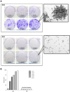
Colony formation escalation in a p53 mutant cell line. a G53 colony formation (crystal violet stain) increases across all cell seeding densities (50 to 600 cells) over 14 days compared to G52 (inset magnification × 20). b Colonies were quantified using a stereomicroscope and colony counting pen. G53 colony counts consistently increase over time with increasing cell density, whereas G52 demonstrates no change in formation
Fig. 5
Fig. 5
Higher cell proliferation in p53 mutant. a Labelling index for Ki-67 in G52 and G53 (×40 magnification). b LI was measured as the percentage of positive cells per 1000 cells. *p < 0.05

Similar articles

Cited by

References

    1. Stupp R, et al. Radiotherapy plus concomitant and adjuvant temozolomide for glioblastoma. N Engl J Med. 2005;352(10):987–996. doi: 10.1056/NEJMoa043330. - DOI - PubMed
    1. Lasocki A, et al. Multifocal and multicentric glioblastoma: improved characterisation with FLAIR imaging and prognostic implications. J Clin Neurosci. 2016;31:92–98. doi: 10.1016/j.jocn.2016.02.022. - DOI - PubMed
    1. Paulsson AK, et al. Comparison of clinical outcomes and genomic characteristics of single focus and multifocal glioblastoma. J Neurooncol. 2014;119(2):429–435. doi: 10.1007/s11060-014-1515-1. - DOI - PMC - PubMed
    1. Thomas RP, et al. The incidence and significance of multiple lesions in glioblastoma. J Neurooncol. 2013;112(1):91–97. doi: 10.1007/s11060-012-1030-1. - DOI - PubMed
    1. Hassaneen W, et al. Multiple craniotomies in the management of multifocal and multicentric glioblastoma. Clinical article. J Neurosurg. 2011;114(3):576–584. doi: 10.3171/2010.6.JNS091326. - DOI - PubMed