

A pin detected by ultrasonography within the normal appendix: prior to surgery, an impressive use of ultrasonography to localize an ingested foreign body exactly
- PMID: 32002897
- PMCID: PMC8572297
- DOI: 10.1007/s40477-020-00431-4
A pin detected by ultrasonography within the normal appendix: prior to surgery, an impressive use of ultrasonography to localize an ingested foreign body exactly
Abstract
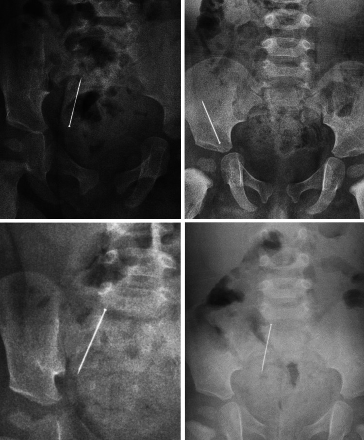
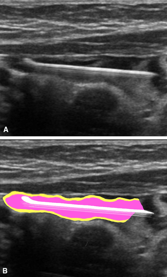
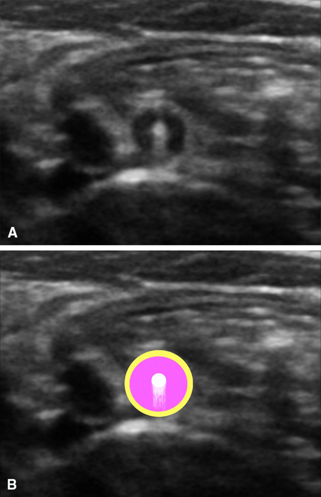
We present a 1-year-old boy who was asymptomatic and brought to the emergency room on suspicion of his having swallowed a pin. Confirmation of ingestion of the pin and its passage through the gut was achieved with abdominal radiography. The pin, which was followed with serial abdominal radiographs, was expected to leave the gastrointestinal tract, but was fixed to the right lower quadrant. When the pin had not passed after 10 days, and with increasing concern about the likelihood of perforation, ultrasonography was used to locate its exact position and allow surgical removal. Only a few cases involving the use of ultrasonography to reveal the exact location of an ingested foreign body prior to surgery have been reported ın the literature. This case presents an impressive example of the use of ultrasonography to reveal the intra-appendiceal location of an ingested foreign body, and to facilitate its surgical removal.
Keywords: Appendicitis; Foreign body; Ingestion; Ultrasound.
© 2020. Società Italiana di Ultrasonologia in Medicina e Biologia (SIUMB).
Conflict of interest statement
The authors declare that they have no conflict of interest.
Figures

References
-
- Simkovic D, Hladík P, Lochman P. Unusual cause of the acute appendicitis. Rozhl Chir. 2004;83:365–367. - PubMed
Publication types
MeSH terms
LinkOut - more resources
Full Text Sources
Medical
