. 2020 Jan 29;12(2):1685-1703.
doi: 10.18632/aging.102707. Epub 2020 Jan 29.

The Interleukin-33/ST2 axis promotes glioma mesenchymal transition, stemness and TMZ resistance via JNK activation

Affiliations

The Interleukin-33/ST2 axis promotes glioma mesenchymal transition, stemness and TMZ resistance via JNK activation

Lin Lin et al. Aging (Albany NY). .

Abstract

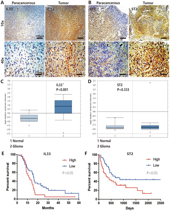
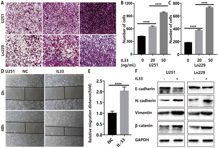
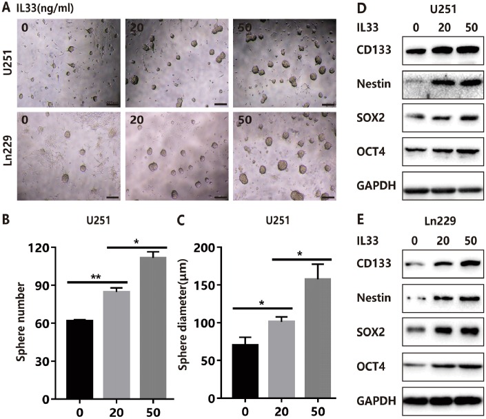
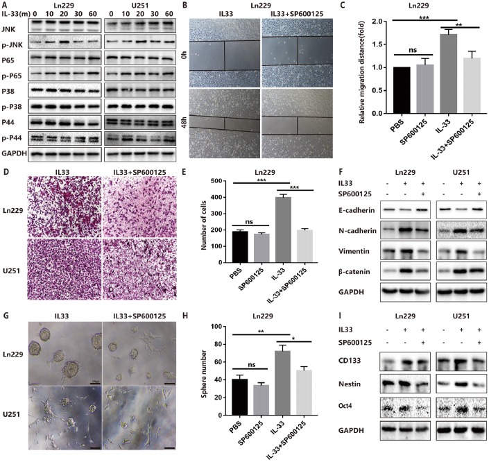
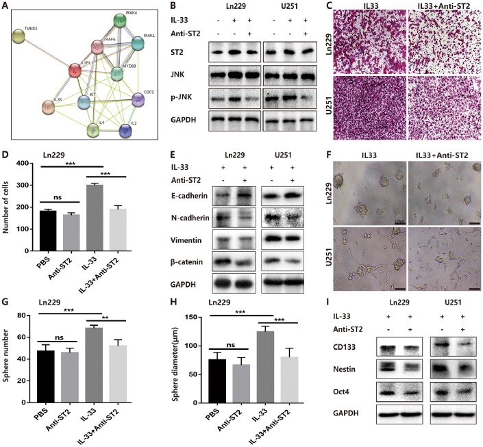
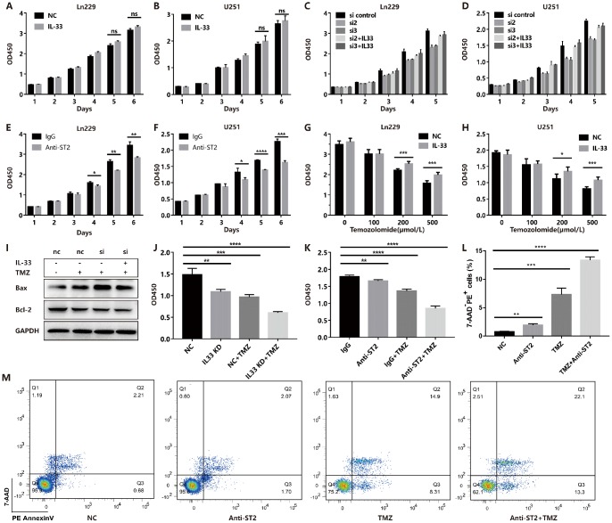
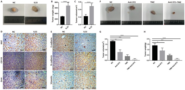
IL-33 is an important member of the IL-1 family which has pleiotropic activities in innate and adaptive immune responses. Recently, some researchers have focused on the function of cellular immunity in the development of tumor. The biological role of IL-33 in glioma is poorly understood. In this study, we showed that glioma cells and tissues expressed higher levels of IL-33 and its receptor ST2 compared to normal brain. Clinically, IL-33 expression was associated with poor survival in patients with glioma. Administration of human IL-33 enhanced cell migration, invasion, epithelial to mesenchymal transition and stemness. Anti-ST2 blocked these effects of IL-33 on tumor. Mechanistically, IL-33 activated JNK signaling pathway via ST2 and increased the expression of key transcription factors that controlled the process of EMT and stemness. Moreover, IL-33 prevented temozolomide induced tumor apoptosis. Anti-ST2 or knockdown IL-33 increased the sensitivity of tumor to temozolomide. Thus, targeting the IL-33/ST2 axis may offer an opportunity to the treatment of glioma patients.

Keywords: EMT; IL-33; glioma; stemness; temozolomide.


Conflict of interest statement

CONFLICTS OF INTEREST: The authors declare that they have no conflicts of interest.

Figures

Figure 1
Figure 1
IL-33 and ST2 expression was increased in glioma and correlated with patient prognosis. (A and B) IL-33 and ST2 expression was detected with conventional immunohistochemical staining in clinical glioma samples. The representative images showed IL-33 or ST2 expression was increased in tumor tissues compared with paracancerous tissues. (C and D) The mRNA expression data of glioma compared with normal brain tissues in the TCGA Database (n=552), the expression of IL-33 was significantly increased in tumor tissues (p<0.001) while ST2 was upregulated moderately with no statistical significance (p=0.333). (E and F) The association between the survival in patients with glioma and IL-33/ST2 expression (n=66 and n=74 for (E and F) respectively). Survival functions were estimated by Kaplan–Meier methods. Hazard ratios (HR) for (E) High/Low=1.921, Low/High=0.575; HR for (F) High/Low=1.828, Low/High=0.547.
Figure 2
Figure 2
IL-33 promotes glioma cell invasion, migration and mesenchymal transition. (A) Effects of IL-33 on glioma cells invasion. Glioma cell lines U251 and Ln229 were subject to transwell assay for 24 hours. IL-33 was added in different concentrations. (B, C) The number of cells moved through the chambers were counted for per field of view and analyzed. Each column represents three independent experiments; Results are expressed as the mean±SD; n=3; ***, P < 0.001; ****, P < 0.0001. (DE) IL-33 promotes the migration of glioma cells U251 by wound healing assay. The tumor cells moving distance was detected and divided by control group as relative migration distance for statistical analysis. Results are expressed as the mean±SD; n=3; ****, P < 0.0001. (F) The levels of core epithelial marker E-cadherin and mesenchymal markers including vimentin, N-cadherin and β-catenin were detected by Western blotting.
Figure 3
Figure 3
IL-33 promotes glioma stemness. (A) Effects of IL-33 on glioma cells stemness. Glioma cell lines U251 and Ln229 were subject to sphere formation assay for 7 days. IL-33 was added in different concentrations. Representative images of spheres of glioma cells are shown. Scale bar, 200 μm. (B and C) The mean numbers and diameters of spheres were counted and analyzed. Each column represents three independent experiments; Results are expressed as the mean±SD; n=3; *, P < 0.05; **, P < 0.01. (D and E) The levels of core stem cell genes including CD133, Nestin, Sox2 and Oct4 were detected by Western blot.
Figure 4
Figure 4
IL-33 promotes glioma EMT and stemness via JNK activation. (A) Effects of IL-33 on NF-κB and MAPK family signal in glioma cells. The cells were treated with IL-33 (20 ng/mL) in different periods of time. The expression of phosphorylated and total proteins was detected by Western blot. (B) Effects of the JNK inhibitor SP600125 on IL-33 induced migration by Wound healing assay. The cells were treated with IL-33 (20 ng/mL) and/or SP600125 (10 μg/mL) for 48 hours. (C) The tumor cells moving distance was detected. Experimental group (IL-33, SP600125 and IL-33+SP600125)/Control group were calculated for statistical analysis. Results are expressed as the mean ± SD; n=3; **, P < 0.01; ***, P < 0.001. (D) Transwell assay indicated the effects of SP600125 on IL-33 induced invasion. (E) The cells moved through the chambers from four groups (PBS, SP600125, IL-33 and IL-33+SP600125) were counted and analyzed. Results are expressed as the mean ± SD; n=3; ***, P < 0.001. (F) Effects of the JNK inhibitor SP600125 on EMT related proteins in glioma cells. N-cadherin, E-cadherin, Vimentin and β-catenin proteins were detected by Western blotting. (G) Effects of JNK inhibitor on glioma cells stemness. Glioma cell lines were subject to sphere formation assay. Representative images of spheres of glioma cells are shown. Scale bar, 100 μm. (H) Sphere number from four groups were counted and analyzed. Results are expressed as the mean ± SD; n=3; *, P < 0.05; **, P < 0.01. (I) The expression of CD133, Nestin and Oct4 were detected by Western blot.
Figure 5
Figure 5
IL-33 interacts with ST2 to activate JNK-enhanced invasion, EMT and stemness. (A) IL-33 and ST2 interaction network from string. (B) Effects of IL-33 on ST2 expression and anti-ST2 blocked the IL-33-induced JNK activation by western blot. (C) Effects of anti-ST2 on the role of IL-33-induced glioma invasion. Glioma cell lines were subject for transwell assay. (D) IL-33 (20ng/mL) and/or anti-ST2 antibody (1 ug/mL) were added in the cell culture. Results are expressed as the mean ± SD; n=3; ***, P < 0.001. (E) Effects of anti-ST2 on the role of IL-33-stimulated EMT related protein expression. Glioma cells were cultured with IL-33 (20 ng/mL) and/or anti-ST2 antibody (1 ug/mL) for 48 hours. The expression of N-cadherin, E-cadherin, Vimentin and β-catenin were quantified by western blot. (F) Effects of anti-ST2 on the role of IL-33-induced glioma stemness. Anti-ST2 blocked the IL-33-induced sphere formation. Representative images of spheres of glioma cells are shown. Scale bar, 100 μm. (GH), The mean numbers and diameters of spheres were counted and analyzed. Results are expressed as the mean ± SD; n=3; **, P < 0.01; ***, P < 0.001. (I) The levels of stemness related genes including CD133, Nestin and Oct4 were detected by Western blot. Results are shown as mean of the data from at least three independent experiments.
Figure 6
Figure 6
IL-33 prevents TMZ-induced apoptosis and blocked IL-33/ST2 increases tumor apoptosis. (A and B) Effects of IL-33 on proliferation of glioma. Glioma cell lines were cultured with or without IL-33 (20 ng/mL) for 6 days. The cell viability was determined by CCK-8 assay. (C and D) Effects of si-IL-33 on glioma cells proliferation. Si-IL-33 and control si-RNA expressing glioma cells were cultured with or without IL-33 (20 ng/mL) for 5 days. The cell viability was determined by CCK-8. (E and F) Effects of anti-ST2 on glioma cells proliferation. Glioma cell lines Ln229 and U251 were treated with anti-ST2 antibody (1 ug/mL) or IgG control for 6 days. The cell viability was determined by CCK-8. (G and H) Effects of IL-33 on glioma chemotherapy. Glioma cell lines Ln229 and U251 were cultured with or without IL-33 (20 ng/mL) for 24 hours and were subsequently exposed to TMZ for 48 hours. The cell viability was determined by CCK-8 assay. (IK) IL33 was knocked down by si-IL-33 and si-control, the si and control groups were treated with IL-33 (20 ng/mL) or/and TMZ (200uM). The expression of Bax, Bcl-2 were detected by western blot (I) and the cell viability was determined by CCK-8 (J and K). (LM) We performed the Annexin V/PI staining to measure apoptosis. Glioma cells were treated with PBS, anti-ST2, TMZ and anti-ST2+TMZ. Percent of 7-AAD-PE+ cells were counted for analysis. All results are expressed as the mean ± SD from at least three independent experiments; n=4; *, P < 0.05; **, P<0.01; ***, P<0.001; ****, P<0.0001.
Figure 7
Figure 7
IL-33 promotes glioma tumorigenesis, EMT and stemness in vivo. (A) Glioma cells (Ln229) were subcutaneously injected into nude BALB/c mice in the presence or absence of 1ug/ml IL-33 and allowed to grow until tumors formed (4 weeks). (B and C) The tumor volume and tumor weight were monitored at 4 weeks after injection. Results are expressed as the mean of tumor volume ± SD; n=5; ****, P<0.0001. (D and E) primary glioma sections form mouse models were stained with proliferative maker Ki67, EMT related antibodies (E-cadherin, N-cadherin and Vimentin) and core stem genes (CD133 and Oct4) for IHC assay. (F) Glioma cells were subcutaneously transplanted into back flanks of NSG mice. After transplanted for 3 weeks, PBS, Anti-ST2, TMZ and Anti-ST2+TMZ were administered intraperitoneally (i.p.) into mice for consecutive 7 days. All NSG mice were killed in the sixth week. (G and H) tumor volume and weight were monitored and analyzed. Results are expressed as the mean of tumor volume ± SD; n=5; *, P < 0.05; **, P<0.01; ***, P<0.001; ****, P<0.0001.

Similar articles

Cited by

References

    1. Schmitz J, Owyang A, Oldham E, Song Y, Murphy E, McClanahan TK, Zurawski G, Moshrefi M, Qin J, Li X, Gorman DM, Bazan JF, Kastelein RA. IL-33, an interleukin-1-like cytokine that signals via the IL-1 receptor-related protein ST2 and induces T helper type 2-associated cytokines. Immunity. 2005; 23:479–90. 10.1016/j.immuni.2005.09.015 - DOI - PubMed
    1. Cayrol C, Girard JP. The IL-1-like cytokine IL-33 is inactivated after maturation by caspase-1. Proc Natl Acad Sci USA. 2009; 106:9021–26. 10.1073/pnas.0812690106 - DOI - PMC - PubMed
    1. Dinarello CA. An IL-1 family member requires caspase-1 processing and signals through the ST2 receptor. Immunity. 2005; 23:461–62. 10.1016/j.immuni.2005.10.004 - DOI - PubMed
    1. Mirchandani AS, Salmond RJ, Liew FY. Interleukin-33 and the function of innate lymphoid cells. Trends Immunol. 2012; 33:389–96. 10.1016/j.it.2012.04.005 - DOI - PubMed
    1. Ali S, Huber M, Kollewe C, Bischoff SC, Falk W, Martin MU. IL-1 receptor accessory protein is essential for IL-33-induced activation of T lymphocytes and mast cells. Proc Natl Acad Sci USA. 2007; 104:18660–65. 10.1073/pnas.0705939104 - DOI - PMC - PubMed

Publication types

MeSH terms