Review
. 2020 Apr 26:38:455-485.
doi: 10.1146/annurev-immunol-111319-023800. Epub 2020 Jan 31.

Knocking 'em Dead: Pore-Forming Proteins in Immune Defense

Affiliations
Review

Knocking 'em Dead: Pore-Forming Proteins in Immune Defense

Xing Liu et al. Annu Rev Immunol. .

Abstract

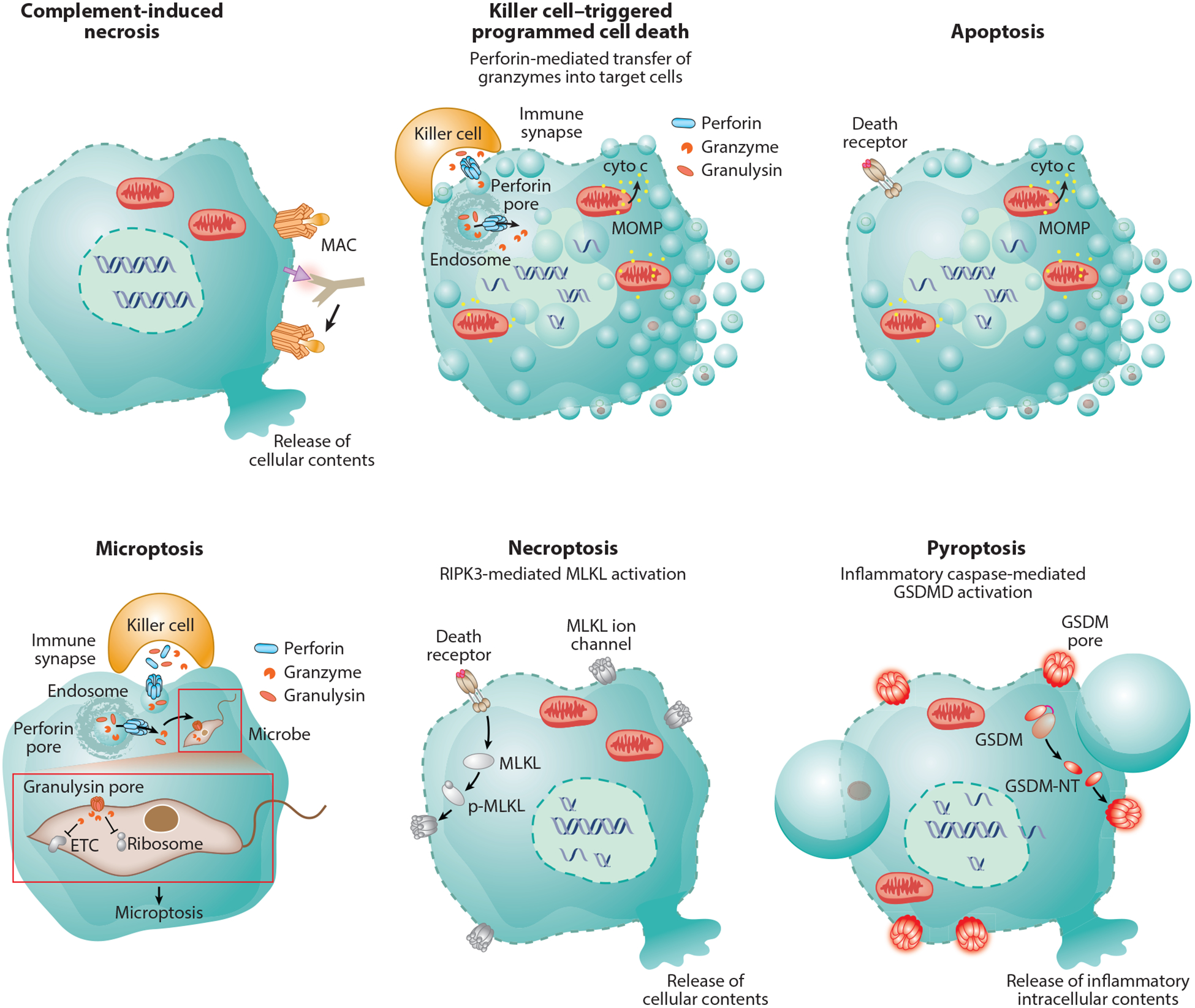
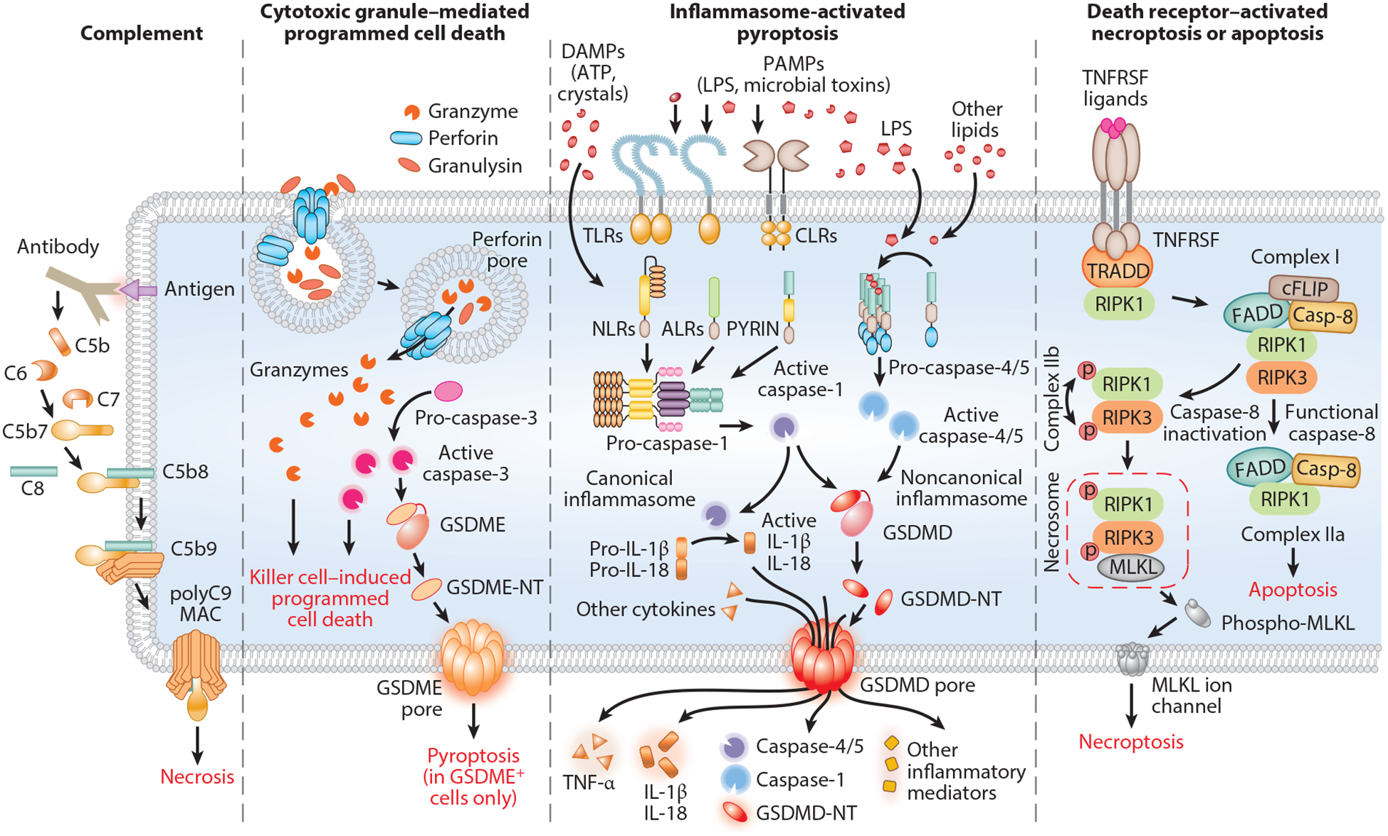
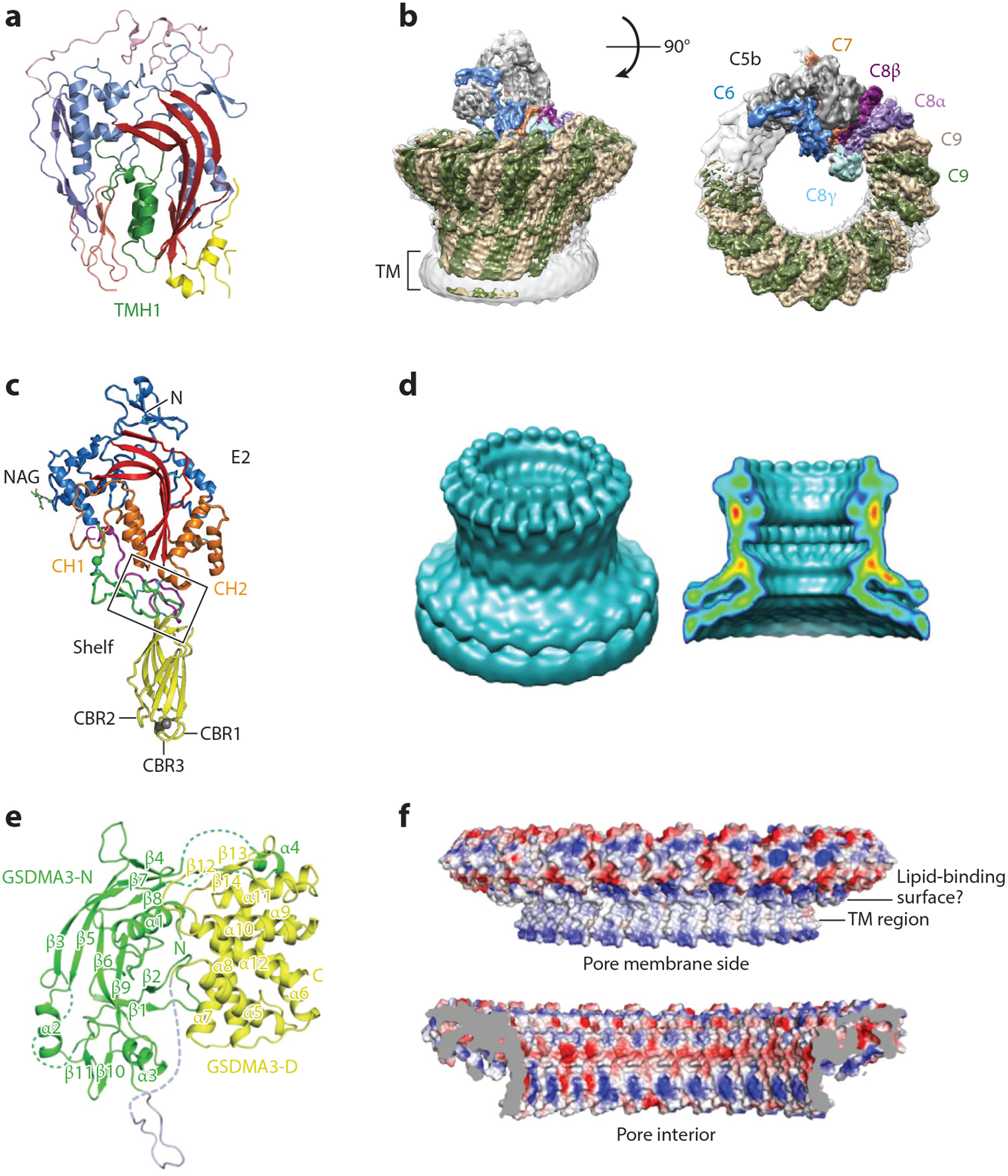
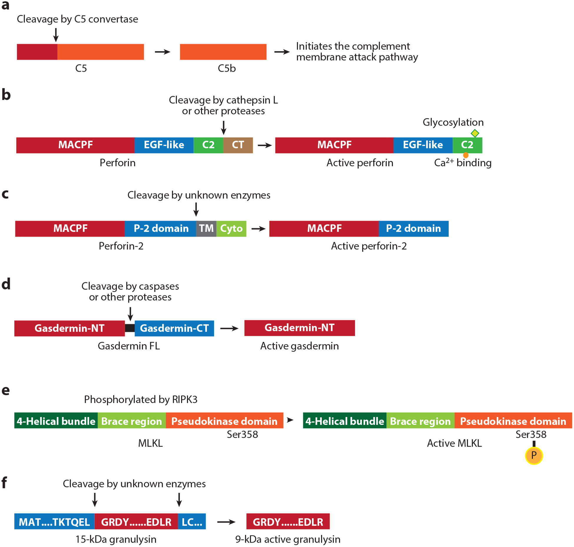
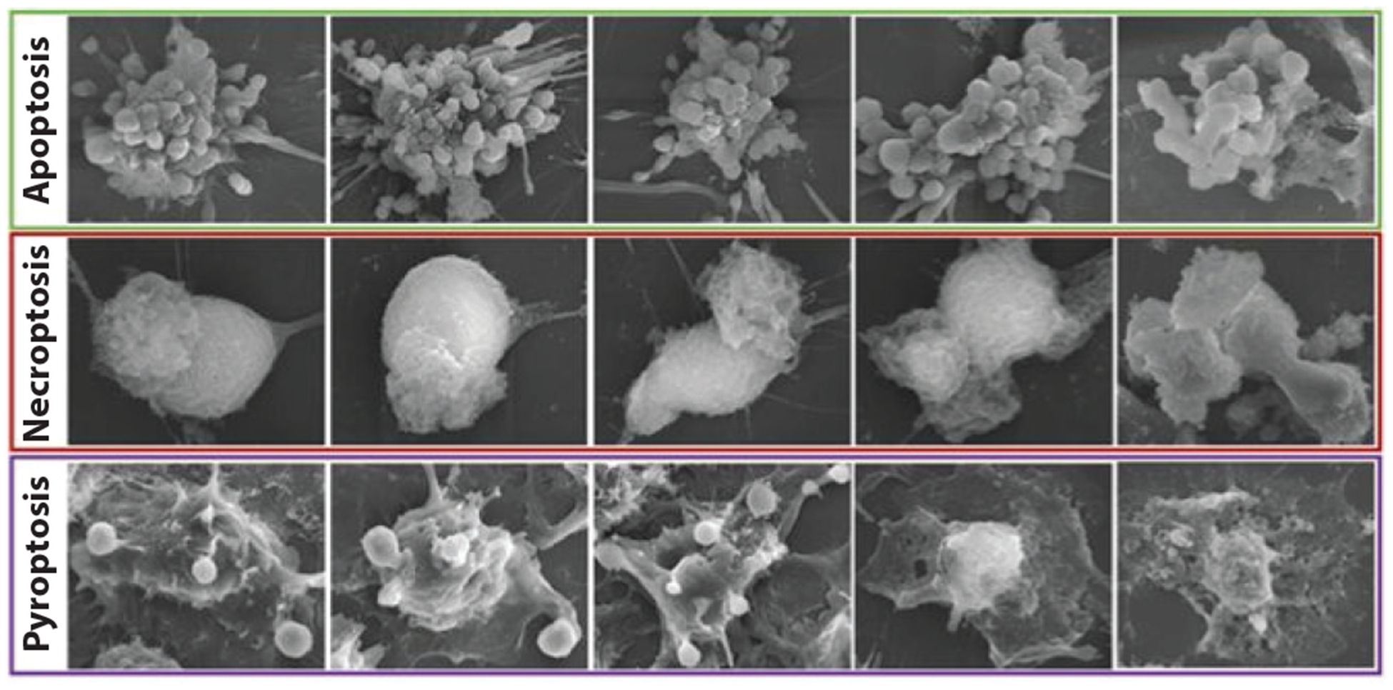
Immune cells use a variety of membrane-disrupting proteins [complement, perforin, perforin-2, granulysin, gasdermins, mixed lineage kinase domain-like pseudokinase (MLKL)] to induce different kinds of death of microbes and host cells, some of which cause inflammation. After activation by proteolytic cleavage or phosphorylation, these proteins oligomerize, bind to membrane lipids, and disrupt membrane integrity. These membrane disruptors play a critical role in both innate and adaptive immunity. Here we review our current knowledge of the functions, specificity, activation, and regulation of membrane-disrupting immune proteins and what is known about the mechanisms behind membrane damage, the structure of the pores they form, how the cells expressing these lethal proteins are protected, and how cells targeted for destruction can sometimes escape death by repairing membrane damage.

Keywords: MLKL; apoptosis; complement; cytotoxic lymphocyte; gasdermin; granulysin; microptosis; necroptosis; necrosis; perforin; pyroptosis.


Figures

Figure 1
Figure 1
Immune membrane damage-mediated cell death. Complement-mediated lysis is a classic example of necrotic cell death characterized by cell membrane rupture. Perforin delivers granzymes to induce programmed target-cell death that resembles apoptosis morphologically with cell shrinkage, membrane blebbing, chromatin condensation, and nuclear fragmentation. Granzyme B also causes apoptosis—it activates caspase-3 and also directly cleaves many caspase-3 substrates to cause mitochondrial outer membrane permeabilization and oligonucleosomal double-stranded DNA breaks, visualized on gels as DNA ladders. Perforin also delivers granulysin into the target cell. Granulysin permeabilizes the cell membrane of intracellular microbes to cause microbial necrosis and allows the granzymes to enter the microbe to proteolytically cause microptosis, programmed cell death of bacteria, fungi, and parasites. Death receptor ligation causes classic apoptosis if caspase-8 or −10 is activated or necroptosis if these caspases are inhibited. In necroptosis, MLKL is phosphorylated, binds to membranes, and perforates them probably by forming ion channels that cause swelling and hypotonic lysis. During pyroptosis, activated in response to invasive pathogens and sterile danger signals, gasdermins are cleaved and the N-terminal fragment forms large plasma membrane pores. Pyroptotic membranes form large ballooning structures, while apoptotic membranes cause small blebs. Because proteins are released through gasdermin pores, the pyroptotic cell likely maintains osmolarity and does not burst. Abbreviations: cyto c, cytochrome C; ETC, electron transport chain; GSDM, gasdermin; GSDM-NT, GSDM N-terminal fragment; GSDMD, gasdermin D; MAC, membrane attack complex; MLKL, mixed lineage kinase domain-like pseudokinase; MOMP, mitochondrial outer membrane permeabilization; p-MLKL, phospho-MLKL.
Figure 2
Figure 2
Molecular mechanisms that activate membrane damage. The complement system is activated by the classic, lectin, or alternative pathway, all of which share a common terminal pathway in which C5 is cleaved into the chemoattractant C5a and the pore-initiating protein C5b. C5b then sequentially binds C6, C7, and C8, which assemble on the target membrane and recruit C9, which oligomerizes to form the MAC pore. Killer cells release cytotoxic granules’ contents (perforin, granzymes, granulysin) into the immune synapse formed with the target cell. Perforin oligomerizes to form short-lived pores in the target cell membrane that trigger membrane repair, which activates endocytosis of the damaged membrane with bound perforin, granzymes, and granulysin. Perforin then forms pores in the endosome, releasing granzymes and granulysin into the target cell cytosol. Cytosolic granzymes induce both caspase-independent and -dependent programmed cell death by cleaving and activating downstream effector proteins. In target cells expressing GSDME, granzyme B-activated caspase-3 can cleave and activate GSDME to convert noninflammatory cell death to inflammatory pyroptosis. In response to cytosolic sensing of microbial infection or danger signals, canonical or noncanonical inflammasomes assemble to activate inflammatory caspases (caspase-1 and caspases-4/−5/−11, respectively). Activated inflammatory caspases cleave GSDMD, releasing its N-terminal fragment, GSDMD-NT, which forms pores in the plasma membrane to cause pyroptosis. Ligand-bound death receptors direct the recruitment and formation of distinct complexes that activate necroptosis or apoptosis. Complex I contains TRADD, RIPK1 and E3 ubiquitin ligases, which inhibit apoptosis by poly-ubiquitinating RIPK1. If RIPK1 is not ubiquitinated, complex IIa activates caspase-8 and initiates apoptosis, whereas when caspase-8 activation is blocked, complex IIb assembles when RIPK1 recruits and phosphorylates RIPK3, which initiates necroptosis by phosphorylating MLKL. Abbreviations: ALR, AIM2-like receptor; CLR, C-type lectin receptor; DAMP, danger-associated molecular pattern; GSDMD, gasdermin D; GSDMD-NT, GSDMD N-terminal fragment; GSDME, gasdermin E; GSDME-NT, GSDME N-terminal fragment; LPS, lipopolysaccharide; MAC, membrane attack complex; MLKL, mixed lineage kinase domain-like pseudokinase; NLR, nucleotide-binding oligomerization domain-like receptor; PAMP, pathogen-associated molecular pattern; TLR, Toll-like receptor; TNFRSF, tumor necrosis factor receptor superfamily; TRADD, TNF-receptor-associated death domain.
Figure 3
Figure 3
Structures of immune pore-forming proteins. (a,c,e) Crystal structures of the monomers of (a) complement C9, (c) perforin, and (e) GSDMA3. (b,d,f) Cryo-electron microscopy reconstruction of the membrane pores formed by (b) the complement membrane attack complex, (d) perforin, and (f) GSDMA3. Abbreviations: GSDMA3, gasdermin A3; TM, transmembrane. Panels a, b, e, and f adapted from References , , , and , respectively; panels c and d adapted from Reference .
Figure 4
Figure 4
Activation of immune membrane-disrupting proteins. (a) The complement cascade is initiated when C5 is processed to generate C5b. (b) Perforin consists of a MACPF domain, an EGF-like domain, and a calcium-binding C2 domain followed by a C-terminal peptide. It is synthesized as an inactive precursor that is glycosylated in its C2 domain and processed in cytotoxic granules by cathepsin L or other unknown proteases. (c) Perforin-2, a membrane-bound protein, consists of a MACPF domain, conserved perforin-2 domain, transmembrane domain and cytoplasmic domain. Its pore-forming activity may be enhanced by proteolytic activation by unknown enzymes to remove the transmembrane domain and release it from vesicular membranes. (d) Gasdermins have N-terminal and C-terminal domains separated by a linker; the C-terminal domain folds back on the N-terminal domain to autoinhibit membrane pore formation. Cleavage in the linker generates the N-terminal pore-forming domain. (e) MLKL, composed of an N-terminal four-helical bundle domain followed by the brace region and a C-terminal pseudokinase inhibitory domain, is activated by phosphorylation of Ser358 by RIPK3. (f) The inactive 15-kDa granulysin propeptide is cleaved after Leu62 and Arg136 by unknown enzymes to produce active granulysin (9 kDa). Abbreviations: CT, C-terminal; Cyto, cytoplasmic; EGF, epidermal growth factor; FL, full length; MACPF, membrane attack complex/perforin; NT, N-terminal; MLKL, mixed lineage kinase domain-like pseudokinase; P-2, perforin-2; TM, transmembrane.
Figure 5
Figure 5
Morphological features of different types of cell death. Adapted from Reference .

References

    1. Rosado CJ, Kondos S, Bull TE, Kuiper MJ, Law RH, et al. 2008. The MACPF/CDC family of pore-forming toxins. Cell Microbiol 10:1765–74 - PMC - PubMed
    1. Dotiwala F, Lieberman J. 2019. Granulysin: killer lymphocyte safeguard against microbes. Curr. Opin. Immunol 60:19–29 - PMC - PubMed
    1. Liu X, Lieberman J. 2017. A mechanistic understanding of pyroptosis: the fiery death triggered by invasive infection. Adv. Immunol 135:81–117 - PMC - PubMed
    1. Shan B, Pan H, Najafov A, Yuan J. 2018. Necroptosis in development and diseases. Genes Dev 32:327–40 - PMC - PubMed
    1. Thiery J, Lieberman J. 2014. Perforin: a key pore-forming protein for immune control of viruses and cancer. Subcell. Biochem 80:197–220 - PubMed

Publication types

MeSH terms

Substances

LinkOut - more resources