. 2020 Jan 31;15(1):e0228226.
doi: 10.1371/journal.pone.0228226. eCollection 2020.

Elemental bioimaging shows mercury and other toxic metals in normal breast tissue and in breast cancers

Affiliations

Elemental bioimaging shows mercury and other toxic metals in normal breast tissue and in breast cancers

Roger Pamphlett et al. PLoS One. .

Abstract

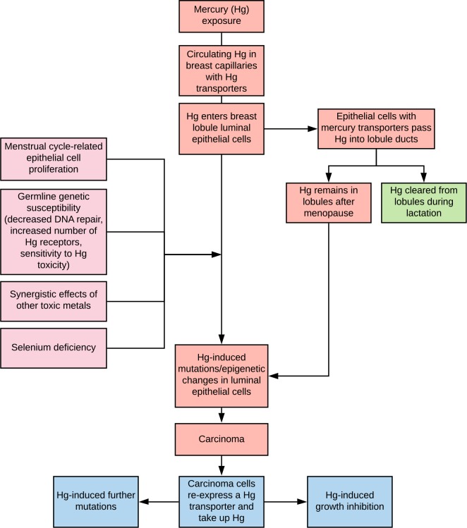
Objective: Exposure to toxic metals such as mercury has been proposed to be a risk factor for the development of breast cancer since some metals can promote genetic mutations and epigenetic changes. We sought to find what toxic metals are present in normal breast tissue and in the tumours of women who had mastectomies for invasive ductal breast carcinoma.

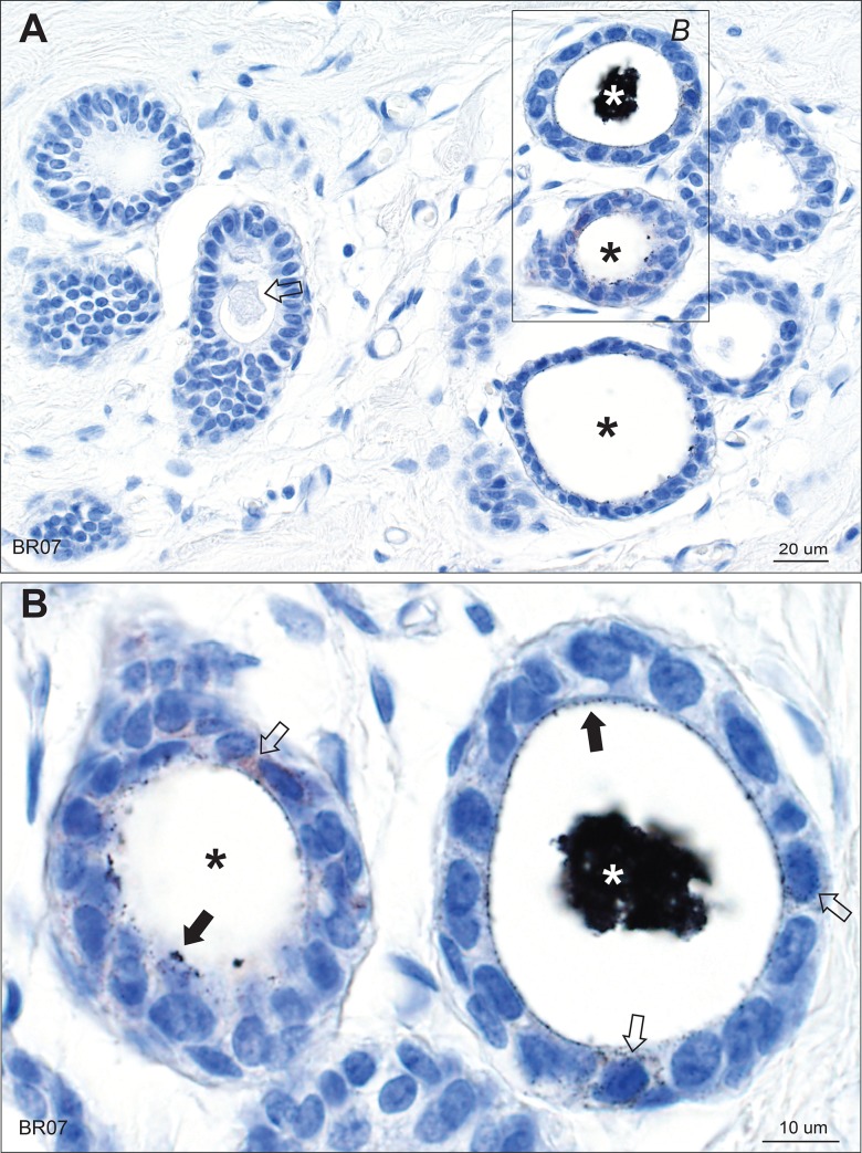
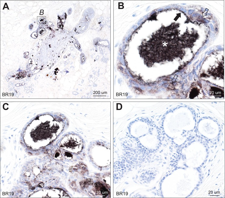
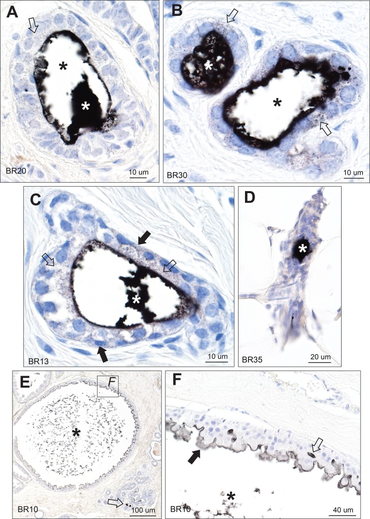
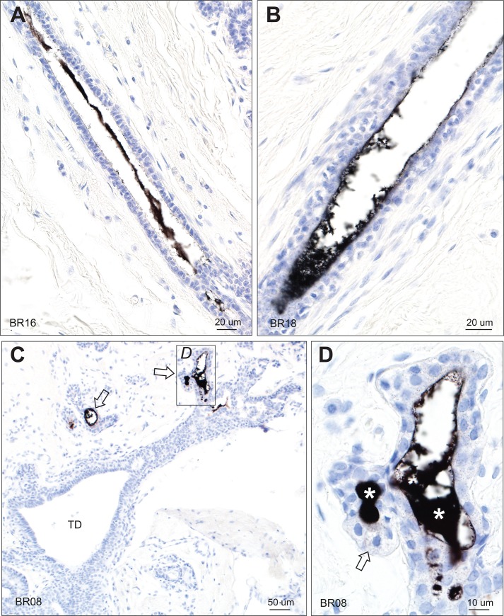
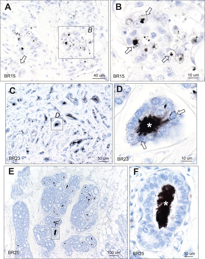
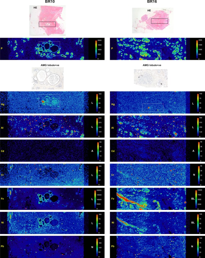
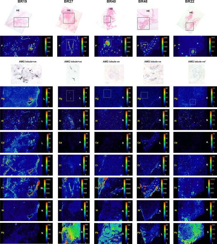
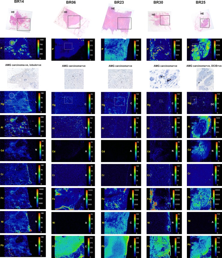
Materials and methods: Formalin-fixed paraffin-embedded blocks from mastectomies for breast carcinoma were examined from 50 women aged 34-69 years. Paraffin blocks selected for elemental analysis were from breast tissue not involved by carcinoma and from the carcinoma itself. Seven micrometer-thick sections were stained with autometallography to demonstrate the presence of mercury, and subjected to laser ablation-inductively coupled plasma-mass spectrometry (LA-ICP-MS) to confirm the presence of mercury and to detect other toxic metals.

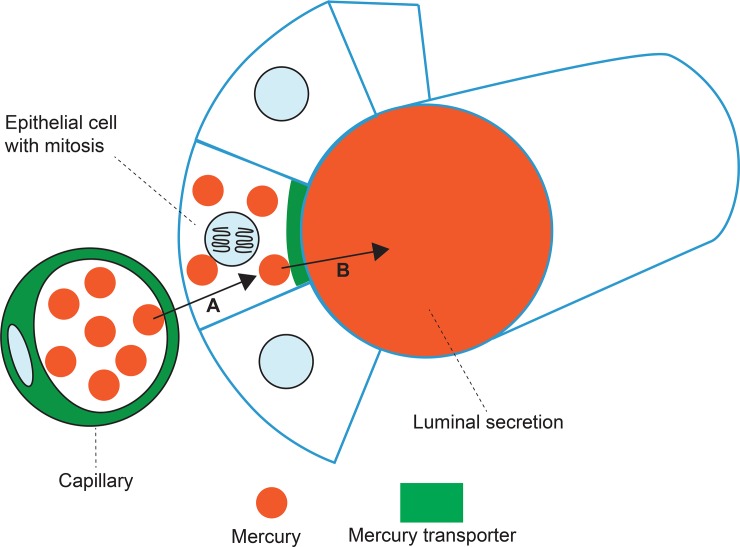
Results: Autometallography-detected mercury was seen in intraductal secretions and some luminal epithelial cells of normal breast lobules in 26 (55%) of the 47 samples where lobules were present, and in 10 (23%) of carcinomas from the 44 samples where carcinoma was present. In eight samples ductal carcinoma in situ was present and one of these contained mercury. LA-ICP-MS confirmed the presence of mercury in samples that stained with autometallography, and detected lead, iron, nickel, aluminium, chromium and cadmium in some samples.

Conclusions: Mercury was present in normal breast lobules in more than half of mastectomy samples that contained an invasive carcinoma, and in a smaller proportion of carcinomas and ductal carcinomas in situ. Other toxic metals that may interact synergistically with mercury could be detected in some samples. These findings do not provide direct evidence that toxic metals such as mercury play a role in the pathogenesis of breast cancer, but suggest that future molecular biological investigations on the role of toxic metals in breast cancer are warranted.


Conflict of interest statement

The authors have declared that no competing interests exist.

Figures

Fig 1
Fig 1. Mercury in breast lobules.
(A) One lobule in the group on the right shows black mercury-stained secretion (white asterisk), and two (black asterisks) show mercury on the luminal surface of epithelial cells. The lobules on the left show no mercury, with unstained luminal secretion visible in one (arrow). (B) Enlarged image of the box in A. The lobule on the right has mercury in its artefactually-shrunken luminal secretion (white asterisk). Some mercury-stained secretion is still attached to the luminal surface of the epithelial cells (solid arrows). Fine particulate mercury staining is present in some epithelial cells (open arrows). In the lobule on the left (black asterisk) much of the secretion has fallen out during processing, but mercury-stained secretion remains attached to the luminal surface of some epithelial cells (closed arrow) and is also present in some epithelial cells (open arrow). AMG/hematoxylin. BR: sample identity number.
Fig 2
Fig 2. Mercury in breast lobules.
(A) Mercury is present in the lumen of many, but not all, lobules. AMG/hematoxylin. (B) Magnified view of the box in A shows mercury within the shrunken luminal secretion (asterisk) of this lobule, with some remaining mercury on the luminal surface of the epithelial cells (solid arrow), and finely dispersed mercury within epithelial cells (open arrows). AMG/hematoxylin. (C, D) The black mercury staining in multiple lumens and cells of these lobules (C) in this AMG/hematoxylin section can be compared to the absence of black staining in a nearby section stained with hematoxylin only (D). BR: sample identity number.
Fig 3
Fig 3. Mercury in breast lobules.
(A, B, C) Mercury is present in the luminal secretion (white asterisks) and epithelial cells (open arrows) of these lobules from three different tissue samples. In A and B, much of the luminal secretion has artefactually dropped out (black asterisks). In C, some mercury may also be present in the cytoplasm of myoepithelial cells (solid arrows) as well as in epithelial cells (open arrows). (D) Mercury is present in the luminal secretion (asterisk) of this atrophic lobule, with surrounding fatty tissue. (E) A large lobule with apocrine metaplasia is filled with fragmented secretion that stains for mercury (asterisk). Some adjacent normal-sized lobules contain mercury in their lumens (arrow). (F) Enlarged view of the box in E shows mercury in the remaining luminal secretion (asterisk), on the luminal surface of apocrine cells (solid arrow), and between apocrine cells (open arrow). AMG/hematoxylin. BR: sample identity number.
Fig 4
Fig 4. Mercury in breast terminal ducts and lobules.
(A, B) Mercury is present within the luminal secretion of these extralobular terminal ducts, but not within the luminal epithelial cells. (C) No mercury is seen in the intralobular terminal duct (TD) of this breast, despite mercury being present in adjacent lobules (arrows). (D) Enlarged view of the box in C shows mercury in the luminal secretion (asterisks) and as fine black particles in epithelial cells (eg, arrow) of this lobule. AMG/hematoxylin. BR: sample identity number.
Fig 5
Fig 5. Mercury in breast carcinomas and ductal carcinoma in situ.
(A) Mercury is seen within the lumens of carcinoma ducts (eg, within the box), whereas other regions of the carcinoma are mercury-free (eg, arrow). (B) Enlarged view of the box in A shows mercury in carcinoma ducts (eg, arrows). (C) Numerous ducts of this carcinoma contain mercury within their lumens (eg, in box). (D) Enlarged view of the box in C shows mercury in the carcinoma-cell-lined lumen of this duct (asterisk), and in adjacent carcinoma cells, where the mercury is often attached to the cell nuclear membrane (eg, arrows). (E) Ductal carcinoma in situ shows many ducts containing mercury (eg, in box). (F) Enlarged view of the box in E shows mercury within a carcinoma-cell-lined lumen (asterisk), but not within adjacent carcinoma cells. AMG/hematoxylin. BR: sample identity number.
Fig 6
Fig 6. LA-ICP-MS of two AMG-positive breast lobules.
Boxes in the hematoxylin-eosin (HE) stained sections in the upper row show the region of LA-ICP-MS analysis. Phosphorus (P) abundance indicates cellular density. Selected regions stained with autometallography (AMG) are indicated in the LA-ICP-MS mercury (Hg) images in dashed boxes. Element distribution = A: absent, B: blood; L: lobules, N: non-localising, S: stroma. Scale = counts per second (proportional to abundance).
Fig 7
Fig 7. LA-ICP-MS of two AMG-positive and three AMG-negative breast lobules.
Boxes in the hematoxylin-eosin (HE) stained sections in the upper row show the region of LA-ICP-MS analysis. Phosphorus (P) abundance indicates cellular density. Selected regions stained with autometallography (AMG) are indicated in the LA-ICP-MS mercury (Hg) images in dashed boxes. Element distribution = A: absent, B: blood; L: lobules, N: non-localising, S: stroma. Scale = counts per second (proportional to abundance). *Sampled in an AMG-negative region.
Fig 8
Fig 8. LA-ICP-MS of one AMG-negative and four AMG-positive breast carcinomas.
Boxes in the hematoxylin-eosin (HE) stained sections in the upper row show the region of LA-ICP-MS analysis. Phosphorus (P) abundance indicates cellular density. Selected regions stained with autometallography (AMG) are indicated in the LA-ICP-MS mercury (Hg) images in dashed boxes. In the AMG-negative BR14 carcinoma, adjacent lobules (eg, in the dashed box) contain mercury; the carcinoma (eg, in the circle) contains aluminium but no mercury. In BR25 both the DCIS (in the dashed box) and the carcinoma contain mercury; the carcinoma also contains aluminium, iron and lead. Element distribution = A: absent, B: blood; C: carcinoma, D: DCIS, L: lobules, N: non-localising. Scale = counts per second (proportional to abundance).
Fig 9
Fig 9. Proposed transfer of circulating mercury through breast epithelial cells into ductules.
(A) A capillary with mercury transporters transfers circulating mercury into a luminal epithelial cell. (B) An epithelial cell with apical mercury transporters transfers mercury into the lumen of the breast ductule. Luminal epithelial progenitor cells that are undergoing mitoses may be particularly vulnerable to the genotoxic effects of mercury.
Fig 10
Fig 10. Potential pathogenetic role of mercury in breast cancer.
Circulating mercury enters breast epithelial cells and then into the ducts. Predisposing factors for mercury being able to induce cancer-inducing genetic and epigenetic changes include the presence of mitoses in mercury-containing epithelial cells, germline genetic susceptibilities to impaired DNA repair or mercury toxicity, additional toxic metals, and a lack of selenium. Mercury is likely to remain in lobules after menopause, with ongoing susceptibility of breast epithelial cells to neoplastic change. Lactation could decrease the risk of mercury-induced breast cancer by clearing the lobules of mercury-containing luminal secretions. Mercury within carcinoma cells could initiate further mutations, or inhibit cell proliferation by direct cellular toxicity.

Similar articles

Cited by

References

    1. Key TJ, Verkasalo PK, Banks E. Epidemiology of breast cancer. Lancet Oncol. 2001;2: 133–140. 10.1016/S1470-2045(00)00254-0 - DOI - PubMed
    1. Madigan MP, Ziegler RG, Benichou J, Byrne C, Hoover RN. Proportion of breast cancer cases in the United States explained by well-established risk factors. J Natl Cancer Inst. 1995;87: 1681–1685. 10.1093/jnci/87.22.1681 - DOI - PubMed
    1. Johnson-Thompson MC, Guthrie J. Ongoing research to identify environmental risk factors in breast carcinoma. Cancer. 2000;88: 1224–1229. 10.1002/(sici)1097-0142(20000301)88:5+<1224::aid-cncr8>3.3.co;2-8 - DOI - PubMed
    1. Brody JG, Rudel RA. Environmental pollutants and breast cancer. Environ Health Perspect. 2003;111: 1007–1019. 10.1289/ehp.6310 - DOI - PMC - PubMed
    1. Brody JG, Moysich KB, Humblet O, Attfield KR, Beehler GP, Rudel RA. Environmental pollutants and breast cancer: epidemiologic studies. Cancer. 2007;109: 2667–2711. 10.1002/cncr.22655 - DOI - PubMed

Publication types