. 2020 Jan 31;15(1):e0227449.
doi: 10.1371/journal.pone.0227449. eCollection 2020.

Pathologic properties of SOD3 variant R213G in the cardiovascular system through the altered neutrophils function

Affiliations

Pathologic properties of SOD3 variant R213G in the cardiovascular system through the altered neutrophils function

Myung-Ja Kwon et al. PLoS One. .

Abstract

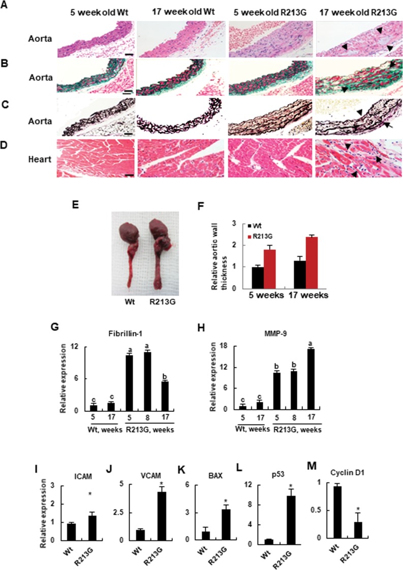
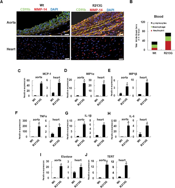
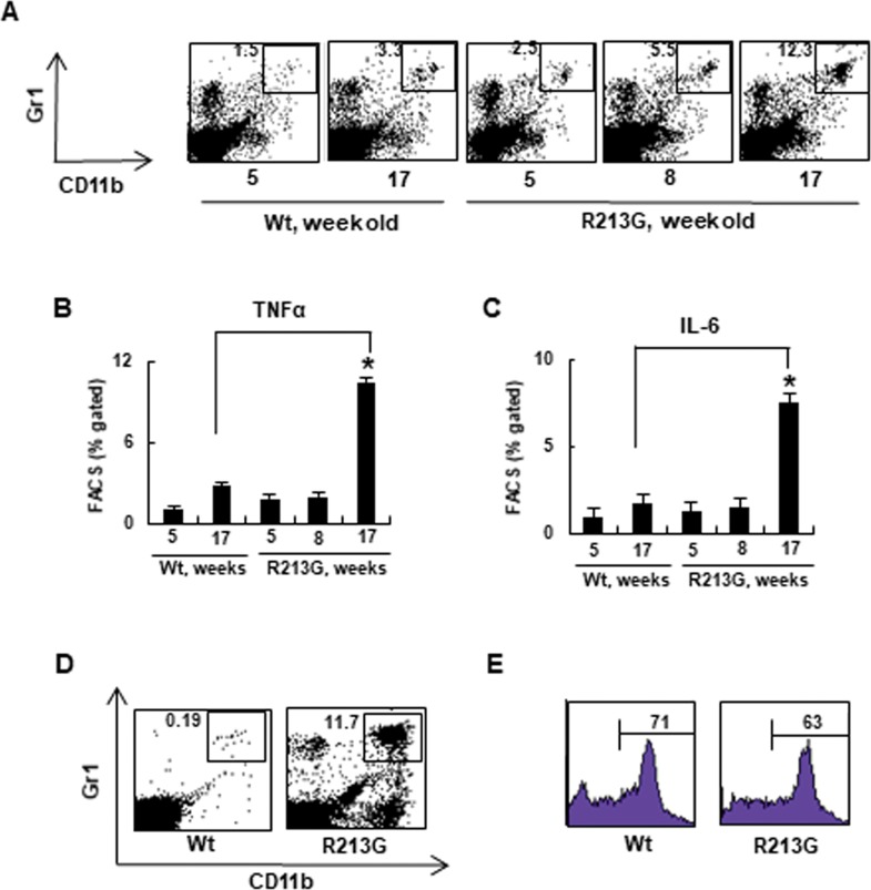
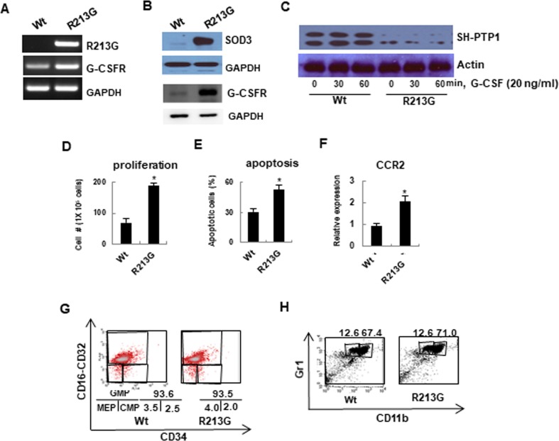
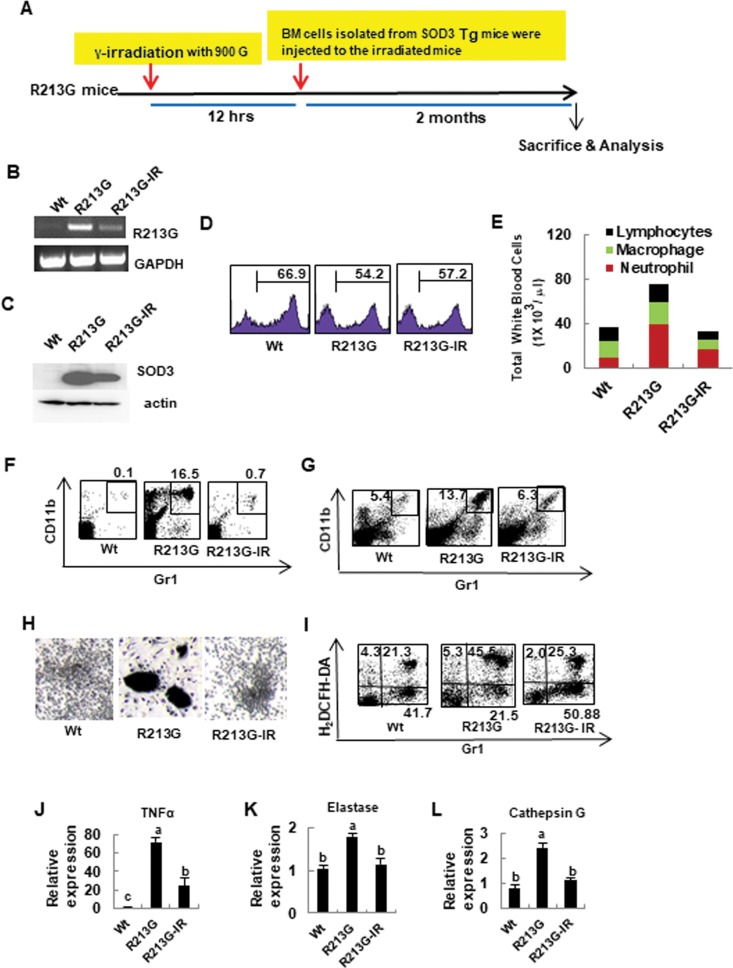
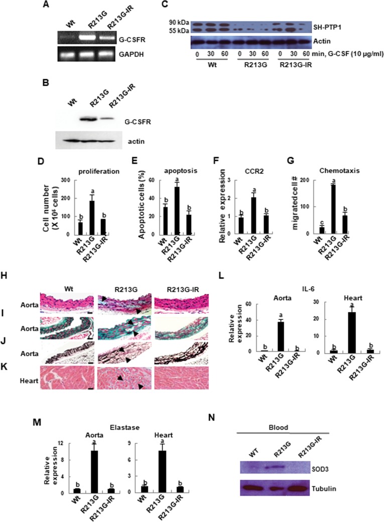
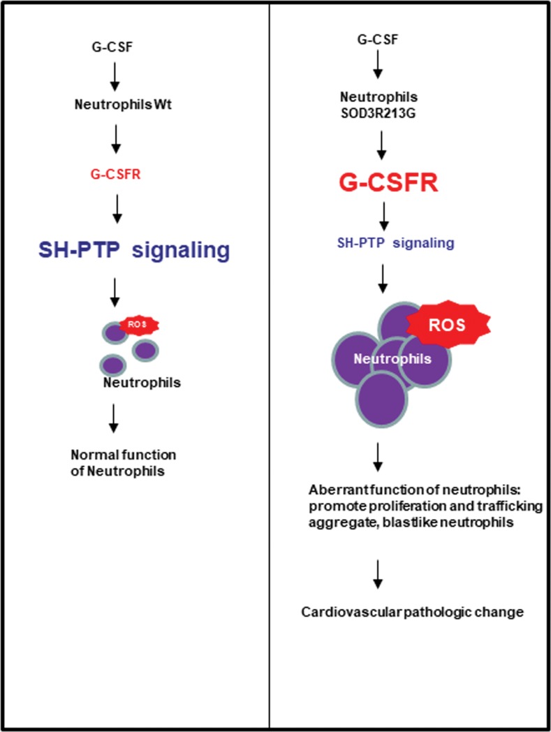
The SOD3 variant, SOD3R213G, results from substitution of arginine to glycine at amino acid 213 (R213G) in its heparin binding domain (HBD) and is a common genetic variant, reported to be associated with ischemic heart disease. However, little is understood about the role of SOD3R213G in innate immune function, and how it leads to dysfunction of the cardiovascular system. We observed pathologic changes in SOD3R213G transgenic (Tg) mice, including cystic medial degeneration of the aorta, heart inflammation, and increased circulating and organ infiltrating neutrophils. Interestingly, SOD3R213G altered the profile of SOD3 interacting proteins in neutrophils in response to G-CSF. Unexpectedly, we found that G-CSF mediated tyrosine phosphatase, SH-PTP1 was down-regulated in the neutrophils of SOD3R213G overexpressing mice. These effects were recovered by reconstitution with Wt SOD3 expressing bone marrow cells. Overall, our study reveals that SOD3R213G plays a crucial role in the function of the cardiovascular system by controlling innate immune response and signaling. These results suggest that reconstitution with SOD3 expressing bone marrow cells may be a therapeutic strategy to treat SOD3R213G mediated diseases.


Conflict of interest statement

The authors have declared that no competing interests exist.

Figures

Fig 1
Fig 1. Aortic degeneration and heart inflammation are presented in SOD3R213G mice.
A-D. Aortic degeneration and heart inflammation in SOD3R213G mice. Abdominal aorta (A-C) and heart (D) of 17 week old SOD3R213G and Wt mice were isolated, along with those of 5 week old SOD3R213G and Wt mice. H & E (A and D), Masson’s trichrome (B), and Van Geison’s staining (C) were performed. Scale bar, 50 μM. Arrow indicates atrophic myocytes (A, 4th panel), fibroblastic proliferation and hydropic degeneration (B, 4th panel), flattened and broken elastic fibers in the aorta (C, 4th panel), and infiltrated inflammatory cells in the heart (D, 4th panel) in the 17 week SOD3R213G mice. E. Image of the aorta. F. Relative aorta wall thickness. Image of the aorta (E) was taken with a digital camera and relative aorta wall thickness (F) was measured. All images are representative of at least of three independent experiments. G-M. Gene expression in the aorta. Fibrillin-1 (G), MMP-9 (H), ICAM (I), VCAM (J), BAX (K), p53 (L), and Cyclin D1 (M). The gene expression was measured by qRT-PCR. All qRT-PCR data were normalized against the mRNA level of the actin gene. The data represent the mean and SE of at least three independent experiments. Statistical analysis was performed by using ANOVA at p<0.05, or t- test at p<0.001, followed by Scheffe’s post hoc test.
Fig 2
Fig 2. Neutrophils are highly infiltrated in the aorta and heart of SOD3R213G mice.
A. Neutrophils infiltration in the aorta and heart of SOD3R213G mice. Abdominal aorta (upper panel) and heart (lower panel) of 17 week old SOD3R213G or Wt mice were isolated, and immunofluorescence staining and confocal analysis were performed as described in Materials and Methods. NIMP-R14: marker for peripheral neutrophils; CD11b: marker for monocytes, neutrophils, granulocytes, and macrophages. Scale bar, 100 μM. B. Profiles of blood cells. Blood was withdrawn from Wt or SOD3R213G mice, followed by Diff-Quick Staining. Inflammatory cells, including neutrophils, macrophages, or lymphocytes, were classified as described in Materials and Methods. C-E. Chemo-attractant expressions F-H. Pro-inflammatory cytokine expressions, I and J. Elastase and TERT expressions in the aorta and heart. Chemoattractants MCP-1 (C), MIP1α (D), MIP1β (E), and proinflammatory cytokines TNFα (F), IL-1β (G), IL-6 (H), and elastase (I), and TERT (J) expression in the aorta and heart of 17 week old SOD3R213G or Wt mice was assessed by qRT PCR. Immunofluorescence data are representative of at least three independent experiments. All qRT-PCR data were analyzed as Fig 1I–1M. Statistical analysis was performed by t- test (*p<0.001).
Fig 3
Fig 3. Premature aging SOD3R213G mice develop neutrophilia.
A. Changes in neutrophils levels of SOD3R213G mice upon aging. Neutrophils (Gr1+CD11b+) in the spleen of SOD3R213G and Wt mice were assessed by FACS analysis as described in Materials and Methods. B-C. Changes in proinflammatory cytokine levels of SOD3R213G mice upon aging. The levels of cytokines, TNFα (B) and IL-6 (C) of SOD3R213G and Wt mice at the indicated time were assessed by flow cytometry as described in Materials and Methods. D-E. Neutrophil content of SOD3R213G. mice. Neutrophils (Gr1+CD11b+) in the blood (D), and Gr+ bone marrow neutrophils (E) of 17 week old SOD3R213G and Wt mice were assessed by FACS analysis as described in Materials and Methods. FACS data are representative of at least three independent experiments. Statistical analysis was performed by t-test (*p<0.001).
Fig 4
Fig 4. SOD3R213G expressing neutrophils exhibit altered G-CSF mediated signaling, promoting proliferation, apoptosis, and trafficking.
A and B. The expression of SOD3R213G and G-CSFR in neutrophils. The expression of the indicated genes was assessed by RT-PCR (A) and immune blot (B) as described in Materials and Methods. C. Down regulation of SH-PTP1 signaling in neutrophils of 17 week old SOD3R213G mice. G-CSF mediated Src tyrosine phosphatase, SH-PTP1 level (C, Top panel) was assessed by treating the neutrophils isolated from 17 week old SOD3R213G or Wt mice with G-CSF (20 ng/ml) at the indicated time. The cells were lysed and subjected to SDS-PAGE, followed by immune blot with an antibody against SH-PTP1. D-F. Proliferation, apoptosis, and chemokine receptor CCR2 expression in neutrophils. The proliferation (D) and apoptosis (E) of neutrophils were measured by treatment with G-CSF (100 ng/ml) for 5 days as described in Materials and Methods. Chemokine receptor CCR2 expression (F) in neutrophils was assessed by qRT-PCR. G and H. Granulopoiesis and maturation of neutrophils were not affected by SOD3R213G. To assess granulopoiesis (G), BM isolated neutrophils from SOD3R213G or Wt mice were subjected to FACS analysis with the indicated antibodies and myeloid progenitor cells were assessed. GMP: granulocyte-monocyte progenitors, CMP: common myeloid precursors, MEP: megakaryocyte-erythrocyte precursors. For the maturation analysis (H), CD11blowGr1high cells for immature and CD11bhighGr1high cells for mature neutrophils were assessed by FACS analysis. RT-PCR, immunoblot, and FACS data are representative of at least three independent experiments. All qRT-PCR data were analyzed as described in Fig 1I–1M. Statistical analysis was performed by t- test (*p<0.001).
Fig 5
Fig 5. Aberrant neutrophils function of SOD3R213G mice is recovered by reconstitution with SOD3 expressing BM cells.
A. The scheme of BM reconstitution. Reconstitution of SOD3 expressing BM cells in SOD3R213G mice was described in Materials and Methods. B. The expression of SOD3R213G. SOD3R213G expression in neutrophils was assessed by RT-PCR as in Fig 4A. C. The protein expression of SOD3. The SOD3 protein expression in the BM neutrophils was assessed as described in materials and methods. D. The level of BM cells in transplanted SOD3R213G mice. Gr1+ BM cells were assessed as described in Materials and Methods. E. Cell profiles in the blood. The cell profiles, including lymphocytes, macrophages, and neutrophils are assessed as described in Materials and Methods. F and G. The contents of neutrophils in the peripheral blood and spleen. Gr1+CD11b+ neutrophils in the blood (F) and spleen (G) were assessed by FACS analysis as described in Fig 3A and 3D. H. Morphological changes of neutrophils. BM cells were isolated from 17 week old SOD3R213G, transplanted, and Wt mice, and treated with G-CSF as described in Materials and Methods. The morphological image was taken by light microscopy. Scale bar, 200 μm. I. Changes in ROS level. ROS generation was assessed by treatment with fMLP (100 nM, 1 hr) in the BM cells, and stained with H2DCFH-DA, 2’,7’-dichlorofluorescin diacetate (5 μM) with Gr1 and analyzed by FACS as described in Materials and Methods. J-L. Gene expressions of neutrophils in SOD3R213G mice. The expression of TNFα (J), elastase (K), and cathepsin G (L) in neutrophils was measured by qRT PCR. RT-PCR and FACS data are representative of at least three independent experiments. All qRT PCR data were analyzed as in Fig 1I–1M. Statistical analysis was performed by using ANOVA at p<0.05, followed by Scheffe’s post hoc test. R213G-IR represents transplanted SOD3R213G mice.
Fig 6
Fig 6. Altered signaling of neutrophils in SOD3R213G mice is recovered by reconstitution with SOD3 expressing BM cells, and aortic degeneration and heart inflammation are recovered.
A and B. G-CSFR expression of neutrophils in SOD3R213G mice was drastically reduced by BM transplantation. G-CSFR gene expression (A) and protein level (B) of neutrophils in Wt, SOD3R213G, and SOD3R213G-IR mice was assessed as described in Fig 4A. C. Altered G-CSF mediated SH-PTP1 signaling of neutrophils of SOD3R213G mice was recovered by BM transplantation. G-CSF mediated SH-PTP1 levels (top panel) in neutrophils of Wt, SOD3R213G, and SOD3R213G-IR mice, were assessed as described in Fig 4C. D-G. Neutrophils function was recovered by BM transplantation. Proliferation (D), apoptosis (E), and chemokine receptor CCR2, expression (F) in neutrophils were measured and analyzed as described in Fig 4D–4F. Chemotaxis (G) of neutrophils was assessed as described in Materials and Methods. H-K. Aortic degeneration and heart inflammation were recovered by transplantation. Abdominal aorta (H-J) and heart (K) of Wt, SOD3R213G, and R213G-IR mice were isolated. H & E (H and K), Masson’s trichrome (I), and Van Grieson staining (J) were performed as described in Fig 1A–1D. Scale bar, 50 μM. L and M. Gene expression of pro-inflammatory molecules. The expression of IL-6 (L) and elastase (M) in the aorta and heart were assessed by qRT-PCR as described in Fig 1I–1M. N. SOD3 level in the blood of SOD3R213G mice was drastically reduced by BM transplantation. Blood isolated from Wt, SOD3R213G, or transplanted mice was lysed and SDS-PAGE was performed, followed immunoblot with indicated antibodies. All images are representative of at least three independent experiments. Each group has three to four Wt, SOD3R213G, or transplanted mice (R213G-IR) mice for an experiment. Statistical analysis was performed by using ANOVA at p<0.05, followed by Scheffe’s post hoc test, grouping a, b, and c.
Fig 7
Fig 7. Proposed mechanism of cardiovascular pathologic change in SOD3R213G mice.
Neutrophils of SOD3R213G mice are dominantly infiltrated in their organs in response to G-CSF, promoting proliferation and trafficking to their organs. Neutrophils of SOD3R213G mice are sensitized, and down-regulated SH-PTP signaling in response to G-CSF may play a major role.

Similar articles

Cited by

References

    1. Marklund SL, Holme E, Hellner L. Superoxide dismutase in extracellular fluids. Clin Chim Acta. 1982;126(1):41–51. Epub 1982/11/24. 10.1016/0009-8981(82)90360-6 . - DOI - PubMed
    1. Folz RJ, Crapo JD. Extracellular superoxide dismutase (SOD3): tissue-specific expression, genomic characterization, and computer-assisted sequence analysis of the human EC SOD gene. Genomics. 1994;22(1):162–71. Epub 1994/07/01. 10.1006/geno.1994.1357 . - DOI - PubMed
    1. Stralin P, Karlsson K, Johansson BO, Marklund SL. The interstitium of the human arterial wall contains very large amounts of extracellular superoxide dismutase. Arterioscler Thromb Vasc Biol. 1995;15(11):2032–6. Epub 1995/11/01. 10.1161/01.atv.15.11.2032 . - DOI - PubMed
    1. Chu Y, Alwahdani A, Iida S, Lund DD, Faraci FM, Heistad DD. Vascular effects of the human extracellular superoxide dismutase R213G variant. Circulation. 2005;112(7):1047–53. Epub 2005/08/10. 10.1161/CIRCULATIONAHA.104.531251 . - DOI - PubMed
    1. Oury TD, Chang LY, Marklund SL, Day BJ, Crapo JD. Immunocytochemical localization of extracellular superoxide dismutase in human lung. Lab Invest. 1994;70(6):889–98. Epub 1994/06/01. . - PubMed

Publication types

MeSH terms

Substances