Review
. 2021 Jan;599(2):453-469.
doi: 10.1113/JP278701. Epub 2020 Feb 18.

AMPA receptor structure and auxiliary subunits

Affiliations
Review

AMPA receptor structure and auxiliary subunits

Aichurok Kamalova et al. J Physiol. 2021 Jan.

Abstract

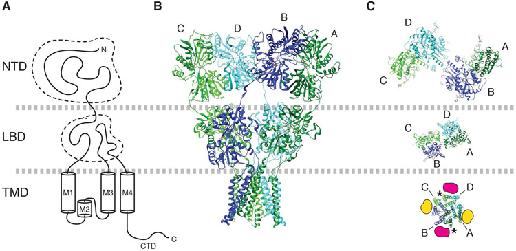
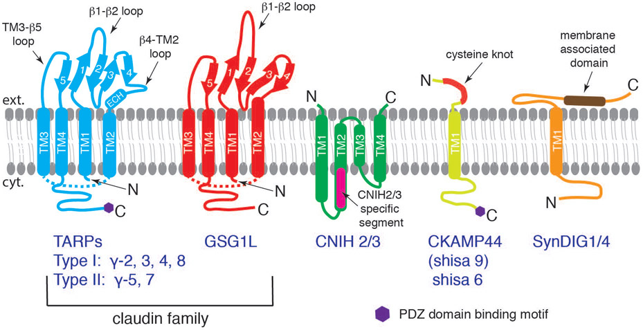
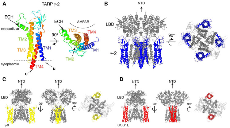
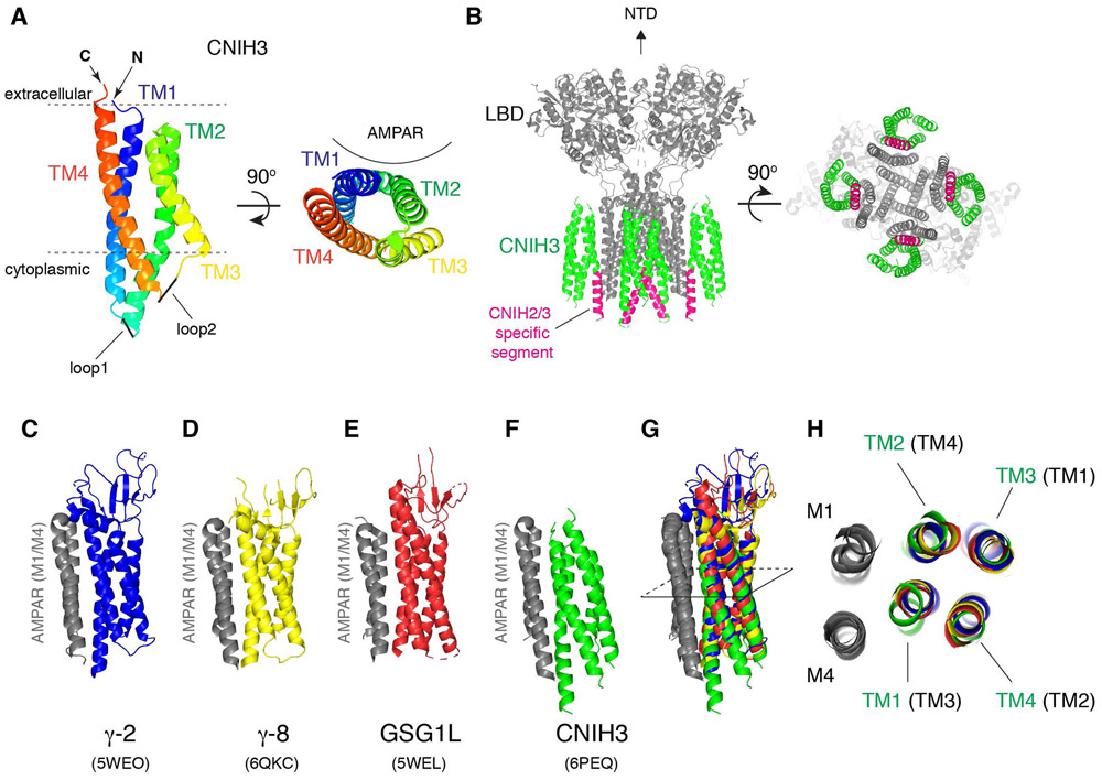
Fast excitatory synaptic transmission in the mammalian brain is largely mediated by AMPA-type ionotropic glutamate receptors (AMPARs), which are activated by the neurotransmitter glutamate. In synapses, the function of AMPARs is tuned by their auxiliary subunits, a diverse set of membrane proteins associated with the core pore-forming subunits of the AMPARs. Each auxiliary subunit provides distinct functional modulation of AMPARs, ranging from regulation of trafficking to shaping ion channel gating kinetics. Understanding the molecular mechanism of the function of these complexes is key to decoding synaptic modulation and their global roles in cognitive activities, such as learning and memory. Here, we review the structural and molecular complexity of AMPAR-auxiliary subunit complexes, as well as their functional diversity in different brain regions. We suggest that the recent structural information provides new insights into the molecular mechanisms underlying synaptic functions of AMPAR-auxiliary subunit complexes.

Keywords: AMPA receptors; AMPA type glutamate receptors; CKAMP44; GSG1L; Shisa; SynDIG; TARP; auxiliary subunits; cornichon; cryo-electron microscopy; electrophysiology; ion channel; ion channel gating modulation; ionotropic glutamate receptors; stargazin; structural biology; synaptic plasticity; synaptic transmission.


Conflict of interest statement

Competing interests: The authors have no competing interests and conflict of interests to declare.

Figures

Figure 1.
Figure 1.. Architecture of AMPAR.
A. Domain organization of a GluA subunit of AMPAR. B. Architecture of tetrameric assembly of GluA subunits in the canonical ‘Y’ shape. Subunits are labeled as A (dark green), B (blue), C (green), and D (cyan). PDB model 3KG2 (Sobolevsky et al., 2009) is displayed. C. The NTD, LBD, and TMD layers are viewed from top. The dimer pairs formed at NTD and LBD layer are different; A/B and C/D at NTD, whereas A/D and B/C at LBD. The TMD is pseudo four-fold symmetric. Labels A-D point to the M4 helix of each subunit. In the TMD, the A/B surface is equivalent to the C/D surface (magenta). Similarly, the B/D surface and A/D surface are equivalent (yellow). The locations indicated by magenta and yellow are the binding site for TARPs and CNIH3. The asterisk indicates the locations of lipids found in AMPAR/TARP γ-8 complex (Herguedas et al., 2019).
Figure 2.
Figure 2.. The variety of global conformations sampled by the NTD layer.
The NTD and LBD are connected by a relatively flexible linker. As a result the NTD layer is positioned differently depending on the length of the linker and subunit composition. The GluA subunits are color coded in red, blue, yellow, and green. A. Y-shape global architecture of GluA2cryst homotetramer (PDB: 3KG2). B. GluA2/GluA3 heterotetramer (PDB: 5IDE). C. GluA2 homotetramer in complex with CNIH3 adopting asymmetric NTD layer (PDB: 6U6I and 6UD4). D. Same as C, but adopting pseudo-symmetric NTD layer (PDB: 6U5S and 6UD8). The gap between the NTD and LBD layer is greater in D than in A. In A, the linker is engineered and shorter than wild type. Similar gap was also observed in GluA2 homotetramer without auxiliary subunits (Meyerson et al., 2014).
Figure 3.
Figure 3.. Schematic illustrations of structurally unrelated AMPAR auxiliary subunits.
TARPs (Type I: γ-2, 3, 4, 8 with a canonical PDZ binding motif; Type II: γ-5, 7 with an atypical PDZ binding motif) and GSG1L belong to the claudin superfamily. The α-helices (thick tubes) and β-strands (thick arrows) are depicted in the topology diagram. Both subclasses contain four transmembrane domains, two extracellular loops, and a cytoplasmic C-terminal tail. The β1-β2 loop is significantly longer in GSG1L. TM1~TM4 fold as a helical bundle, and therefore, the cytoplasmic TM2-TM3 loops of TARP and GSG1L (dashed lines) are shorter than how they are illustrated. CNIH is also a four-pass transmembrane protein, which is mostly embedded within the membrane. The N- and C-termini of CNIH are both extracellular. The CNIH2/3 specific segment, absent in CNIH1/4, is indicated (magenta). CKAMP44 (Shisa9) and Shisa6 have one transmembrane domain with an extracellular cysteine-knot motif and a long intracellular C-terminal tail with type II PDZ binding motif. SynDIG1 and 4 have one transmembrane domain and a long extracellular domain containing a membrane-associated domain.
Figure 4.
Figure 4.. Architecture of TARPs and AMPAR/TARP complexes.
A. A ribbon diagram of TARP γ-2 shows the 3D arrangement of the 4-helix bundle made of TM1-4. An extracellular helix (ECH) is attached to TM2. TARP γ-2 from PDB: 5WEO is displayed at two different views, one parallel to the membrane (left) and another from the extracellular side (right). Location of AMPAR is indicated. B. A ribbon diagram of the AMPAR(GluA2)/TARP γ-2 complex. GluA2(gray):TARP γ-2(blue) =4:4 stoichiometry. 5WEO is displayed at two different views (side and bottom), but the NTD was removed for clarity. C. A ribbon diagram of the heteromeric AMPAR(GluA1 and 2)/TARP γ-8 complex. GluA1(gray):GluA2(gray):TARP γ-8(yellow) =2:2:2 stoichiometry. 6QKC is displayed at three different views (side and bottom), but the NTD was removed for clarity. D. A ribbon diagram of the AMPAR(GluA2)/GSG1L complex. GluA2(gray):GSG1L(red)=4:2 stoichiometry. 5WEL is displayed at three different views (side and bottom), but the NTD was removed for clarity.
Figure 5.
Figure 5.. Architecture of CNIH3 and AMPAR/CNIH3 complex.
A. A ribbon diagram of CNIH3 shows the 3D arrangement of the 4-helix bundle made of TM1-4. CNIH3 from PDB: 6PEQ is displayed at two different views, one parallel to the membrane (left) and another from the extracellular side (right). Location of AMPAR is indicated. B. A ribbon diagram of an AMPAR/CNIH3 complex. GluA2(gray):CNIH3(green)=4:4 stoichiometry. 6PEQ is displayed at two different views (side and bottom), but the NTD was removed for clarity. The CNIH2/3 specific segment is highlighted in magenta. C and F. The ribbon diagram of the M1 and M4 of AMPAR (gray) bound to various auxiliary subunits. The binding interface is viewed from the side in parallel to the membrane. C=TARP γ-2, D= TARP γ-8, E= GSG1L, F=CNIH3. G. Superposition of the structures in C-F. Specifically, when the M4 of AMPAR in C-F is aligned, the M1 and the transmembrane helices of the auxiliary subunits superimpose. The M1/M4 of AMPAR form the auxiliary subunit binding module for TARPs, GSG1L, and CNIH3, which are geometrically conserved. H. Cross section at the slice shown in G is viewed from the top. The TM3 and TM4 (of TARP and GSG1L) interface with M1 and M4 of adjacent subunit of AMPAR. In contrast, the TM1 and TM2 of CNIH3 interface with M1 and M4. The TM1 to TM4 of CNIH3 are labeled in green, whereas the corresponding TM in TARPs and GSG1L are labeled in black and in brackets.
Figure 6.
Figure 6.. Intolerance of AMPAR complexes to missense mutations.
Missense tolerance ratio (MTR) is an estimate of the extent of purifying selection or the removal of deleterious alleles (Traynelis et al., 2017). An MTR=1 indicates neutrality and the ratios below the 10th percentile (in red) are under purifying selection. AMPAR auxiliary subunits have varying degrees of tolerance for missense mutations (i.e. low MTR scores). A. GRIA2, which encodes GluA2 subunit, has regions with MTR=0 (pre-M1 and part of M1), no tolerance to variation. Missense mutations associated with neurodevelopmental disorders (G47E, D302G, P528T, Q/R607G/E, G609R, D611N, A639S, F644L, T646L, V647L) are indicated with red dots (Salpietro et al., 2019). B. GluA3 subunits also has multiple regions with MTR=0, highly intolerant to variation. The locations of mutations associated with cognitive impairment (G833R, M706T, R631S, R450Q) are indicated as red dots (Wu et al., 2007). C. MTR distribution for γ-2 shows multiple regions of high intolerance (in red) including TM2. Missense mutation V143L associated with ID is indicated as a red dot. D. Among TARPs, γ-3 has the highest number regions of intolerance including TM1, TM2, and TM4. E. Cysteine knot domain within CKAM44 is highly intolerable to genetic variation. F. CNIH2 has a stretch of amino acids that are highly intolerable to genetic variation. This region is mapped as the critical AMPAR binding site.

References

    1. Amin JB, Salussolia CL, Chan K, Regan MC, Dai J, Zhou HX, Furukawa H, Bowen ME & Wollmuth LP. (2017). Divergent roles of a peripheral transmembrane segment in AMPA and NMDA receptors. J Gen Physiol 149, 661–680. - PMC - PubMed
    1. Armstrong N & Gouaux E. (2000). Mechanisms for activation and antagonism of an AMPA-sensitive glutamate receptor: crystal structures of the GluR2 ligand binding core. Neuron 28, 165–181. - PubMed
    1. Armstrong N, Sun Y, Chen GQ & Gouaux E. (1998). Structure of a glutamate-receptor ligand-binding core in complex with kainate. Nature 395, 913–917. - PubMed
    1. Ayalon G & Stern-Bach Y. (2001). Functional assembly of AMPA and kainate receptors is mediated by several discrete protein-protein interactions. Neuron 31, 103–113. - PubMed
    1. Azumaya CM, Days EL, Vinson PN, Stauffer S, Sulikowski G, Weaver CD & Nakagawa T. (2017). Screening for AMPA receptor auxiliary subunit specific modulators. PLoS One 12, e0174742. - PMC - PubMed

Publication types