. 2020 Mar 3;28(3):363-370.e3.
doi: 10.1016/j.str.2019.12.002. Epub 2020 Jan 30.

Cryoelectron Microscopy Structure of a Yeast Centromeric Nucleosome at 2.7 Å Resolution

Affiliations

Cryoelectron Microscopy Structure of a Yeast Centromeric Nucleosome at 2.7 Å Resolution

David Migl et al. Structure. .

Abstract

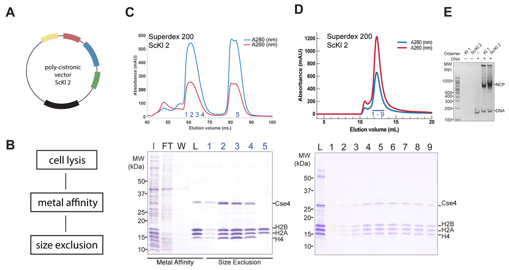
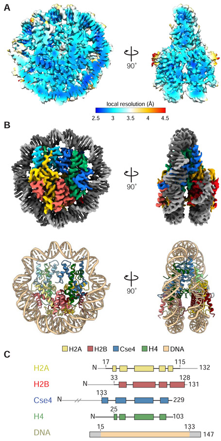
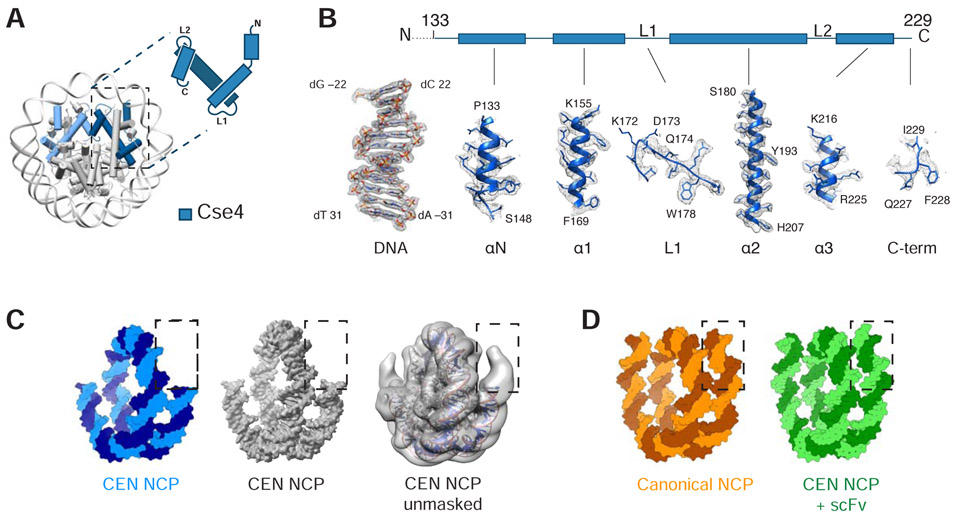
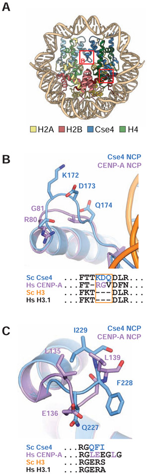
Kinetochores mediate chromosome segregation during cell division. They assemble on centromeric nucleosomes and capture spindle microtubules. In budding yeast, a kinetochore links a single nucleosome, containing the histone variant Cse4CENP-A instead of H3, with a single microtubule. Conservation of most kinetochore components from yeast to metazoans suggests that the yeast kinetochore represents a module of the more complex metazoan arrangements. We describe here a streamlined protocol for reconstituting a yeast centromeric nucleosome and a systematic exploration of cryo-grid preparation. These developments allowed us to obtain a high-resolution cryoelectron microscopy reconstruction. As suggested by previous work, fewer base pairs are in tight association with the histone octamer than there are in canonical nucleosomes. Weak binding of the end DNA sequences may contribute to specific recognition by other inner kinetochore components. The centromeric nucleosome structure and the strategies we describe will facilitate studies of many other aspects of kinetochore assembly and chromatin biochemistry.

Keywords: CENP-A; Cse4; NCP; cryo-EM; histones; kinetochore; nucleosome; structure.


Conflict of interest statement

Declaration of Interests The authors declare no competing interests.

Figures

Figure 1.
Figure 1.
Reconstitution of yeast centromeric nucleosome. A. Schematic representation of poly-cistronic expression vector ScKl 2 representing histone gene order (see details for other expression vectors in Table 1). B. Summary of histone octamer purification workflow. C. Top, size exclusion chromatogram from Superdex 200 column showing elution profile of ScKl 2. Bottom, coomassie-stained 4-12% SDS-PAGE gel with fractions corresponding to metal affinity purification (Input, Flow Through, Wash) and chromatogram on top (Load and numbered fractions). D. Top, size exclusion chromatogram from Superdex 200 column of 601 DNA wrapped centromeric nucleosome expressed from ScKl 2 vector. Bottom, coomassie-stained 4-12% SDS-PAGE gel with fractions corresponding to chromatogram on top. E. Agarose gel showing 601 DNA wrapped centromeric nucleosomes expressed from Kl 1 and ScKl 2 vectors.
Figure 2.
Figure 2.
Cryo-EM structure of yeast centromeric NCP. A. Local resolution map of centromeric NCP based on non-sharpened map. B. Top, cryo-EM density map of centromeric NCP at 2.7Å resolution (unsharpened). Bottom, cartoon representation of centromeric NCP model. (PDB ID: 6UPH) C. Domain representation of histones and 601 Widom DNA where light grey represents regions that have not been built in the model.
Figure 3.
Figure 3.
Structural details of centromeric histone Cse4 and DNA ends. A. Cartoon representation of centromeric (CEN)-NCP and schematic representation of Cse4 histone. B. Density map of centromeric histone Cse4 and a representative DNA region. Dashed line indicates unmodeled N-terminus of Cse4 (residues 1-132). C. Left two, surface representation of 601 DNA from CEN-NCP structure in blue and masked density map of the CEN-NCP at 2.7Å resolution in grey. Right, unmasked density map of the CEN-NCP filtered to low resolution in grey with cartoon representation of the CEN-NCP model (structures from this paper). D. Surface representation of 601 DNA from crystal structure of canonical NCP (PDB ID: 1ID3) and of native apha-satellite DNA from cryo-EM structure of CEN-NCP+scFv (PDB ID: 6E0P) (21, 29).
Figure 4.
Figure 4.
Structural details of Cse4 L1 loop and C-terminus. A. Model of centromeric nucleosome indicating zoomed in regions represented in B and C. B. Overlay of L1 loop from S. cerevisiae Cse4 (blue) and human CENP-A (purple). Sequence alignment of this region is shown below. C. Overlay of S. cerevisiae Cse4 (blue) and human CENP-A (purple) C-terminus with sequence alignment of the region shown below.

References

    1. Adams PD, Afonine PV, Bunkoczi G, Chen VB, Davis IW, Echols N, Headd JJ, Hung LW, Kapral GJ, Grosse-Kunstleve RW, et al. (2010). PHENIX: a comprehensive Python-based system for macromolecular structure solution. Acta Cryst (2010). D66, 213–221. - PMC - PubMed
    1. Anderson M, Huh JH, Ngo T, Lee A, Hernandez G, Pang J, Perkins J, and Dutnall RN (2010). Co-expression as a convenient method for the production and purification of core histones in bacteria. Protein Expression and Purification 72, 194–204. - PubMed
    1. Bilokapic S, Strauss M, and Halic M (2017). Histone octamer rearranges to adapt to DNA unwrapping. Nat Struct Mol Biol 25, 101–108. - PMC - PubMed
    1. Black BE, foltz DR, Chakravarthy S, Luger K, woods VL, and Cleveland DW (2014). Structural determinants for generating centromeric chromatin. 430, 1–5. - PubMed
    1. Cao S, Zhou K, Zhang Z, Luger K, and Straight AF (2018). Constitutive centromere-associated network contacts confer differential stability on CENP-A nucleosomes in vitro and in the cell. Molecular Biology of the Cell 1–12. - PMC - PubMed

Publication types

MeSH terms