. 2020 Mar 3;31(3):518-533.e10.
doi: 10.1016/j.cmet.2020.01.001. Epub 2020 Jan 30.

Macrophage Metabolism of Apoptotic Cell-Derived Arginine Promotes Continual Efferocytosis and Resolution of Injury

Affiliations

Macrophage Metabolism of Apoptotic Cell-Derived Arginine Promotes Continual Efferocytosis and Resolution of Injury

Arif Yurdagul Jr et al. Cell Metab. .

Abstract

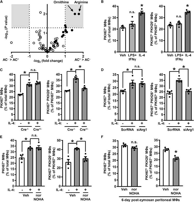
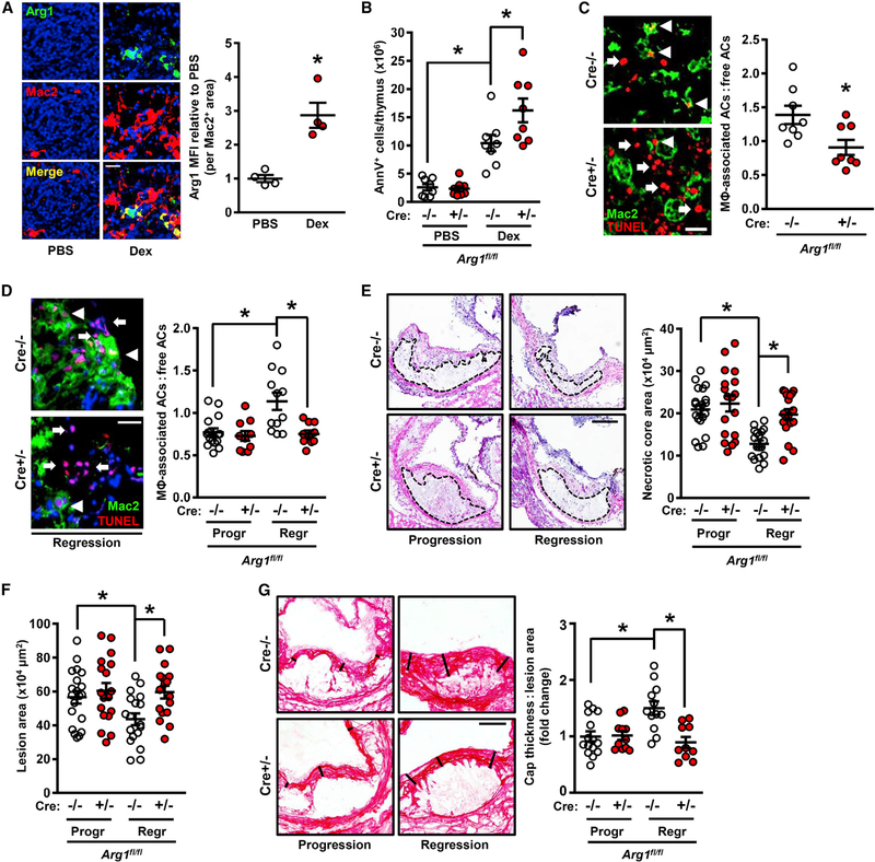
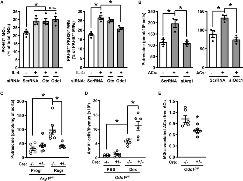
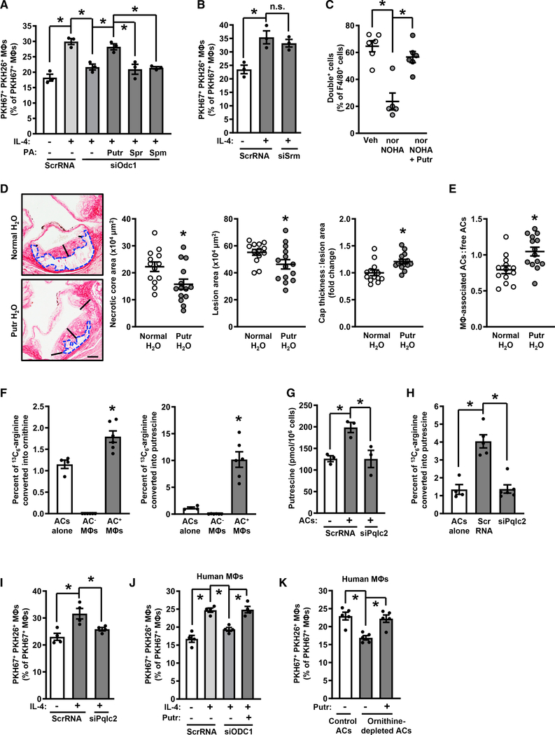
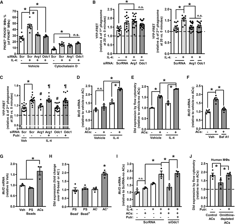
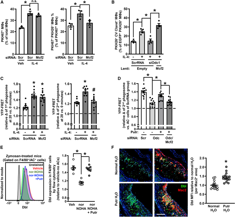
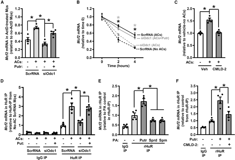
Continual efferocytic clearance of apoptotic cells (ACs) by macrophages prevents necrosis and promotes injury resolution. How continual efferocytosis is promoted is not clear. Here, we show that the process is optimized by linking the metabolism of engulfed cargo from initial efferocytic events to subsequent rounds. We found that continual efferocytosis is enhanced by the metabolism of AC-derived arginine and ornithine to putrescine by macrophage arginase 1 (Arg1) and ornithine decarboxylase (ODC). Putrescine augments HuR-mediated stabilization of the mRNA encoding the GTP-exchange factor Dbl, which activates actin-regulating Rac1 to facilitate subsequent rounds of AC internalization. Inhibition of any step along this pathway after first-AC uptake suppresses second-AC internalization, whereas putrescine addition rescues this defect. Mice lacking myeloid Arg1 or ODC have defects in efferocytosis in vivo and in atherosclerosis regression, while treatment with putrescine promotes atherosclerosis resolution. Thus, macrophage metabolism of AC-derived metabolites allows for optimal continual efferocytosis and resolution of injury.

Keywords: arginase; arginine; atherosclerosis; atherosclerosis regression; efferocytosis; inflammation resolution; intracellular metabolism; macrophage; polyamines; putrescine.


Conflict of interest statement

Declaration of Interests The authors declare no competing interests.

Figures

Figure 1.
Figure 1.. Arg1 Drives Continual Efferocytosis in Resolving-Type Mouse Macrophages
(A) A volcano plot of AC and AC+ mouse bone-marrow-derived macrophages from an efferocytosis assay followed by mass spectrometry analysis for acylcarnitines (open circles) and amino acids (closed circles). Gray boxes show areas of the plot with changes ≥2-fold at p < 0.05. Raw values are shown in Table S1 (n = 3 biological replicates). (B) The percentages of PKH67+ macrophages versus total macrophages (left panel) and PKH67+ PKH26+ macrophages versus PKH67+ macrophages (right panel) as determined by epifluorescence microscopy of macrophages treated with either vehicle (Veh), LPS + IFNγ, or IL-4 in a two-step efferocytosis assay utilizing PKH67-labeled ACs in the first round of efferocytosis and PKH26-labeled ACs in the second round. Illustrative images are shown in Figure S1A. (C) Single- and double-AC efferocytosis (as in B) by macrophages isolated from Arg1fl/fl (Cre−/−) and Arg1fl/fl Lysz2-Cre+/− (Cre+/−) mice and then treated for 24 h with vehicle or IL-4. (D) Single- and double-AC efferocytosis in macrophages transfected with scrambled RNA (ScrRNA) or siArg1 and then treated 24 h with vehicle or IL-4. (E) Single- and double-AC efferocytosis in macrophages that were treated with vehicle or IL-4 and then with vehicle or 500 μM nor-NOHA for 1 h before the addition of the first AC. (F) Single- and double-AC efferocytosis in peritoneal macrophages harvested from mice 6 days after i.p. injection with 0.1 mg of zymosan A and then incubated for 1 h with vehicle or 500 μM nor-NOHA. All values are means ± SEM; *p < 0.05; n.s., not significant; n = 3–4 biological replicates.
Figure 2.
Figure 2.. Myeloid-Arg1 Deletion Impairs Efferocytosis In Vivo
(A) Arg1 immunostaining with quantification of thymic sections from mice 18 h after i.p. injection with PBS or dexamethasone (Dex) (n = 4 mice per group). Scale bar, 50 μm. (B) Annexin V+ cells in thymi from myeloid-Arg1 WT (Cre−/−) and KO (+/−) mice harvested 18 h after injection with PBS or dexamethasone (n = 8 mice per group). (C) Quantification of efferocytosis (macrophage-associated ACs:free ACs ratio) in thymic sections from the mice in (B) (n = 8 per group). Arrowheads and arrows depict macrophage-associated and free TUNEL+ cells, respectively. (D–G) Arg1fl/fl Ldlr−/− (Cre−/−) and Arg1fl/fl Lysz2-Cre+/− Ldlr−/− (Cre+/−) mice placed on atherosclerosis progression (Progr) or regression (Regr) protocols were assayed for the following parameters: (D) efferocytosis (scale bar, 40 μm), (E and F) aortic root (E) necrotic core and (F) lesion areas (n = 17–20 mice per group; scale bar, 200 μm), and (G) collagen cap thickness (n = 10–15 mice per group; scale bar, 100 μm). All values are means ± SEM; *p < 0.05; n.s., not significant.
Figure 3.
Figure 3.. Deletion of Odc1 Causes Defective Continual Efferocytosis
(A) Efferocytosis by ScrRNA-, siOtc-, or siOdc1-transfected macrophages treated with vehicle or IL-4 (n = 4 biological replicates). (B) Putrescine content of IL-4-treated macrophages that were incubated with PKH67-labeled ACs for 45 min and then after 2 h sorted into AC and AC+ populations (n = 3 biological replicates). (C) Putrescine content of aortas from Arg1fl/fl Ldlr−/− (Cre−/−) and Arg1fl/fl Lysz2-Cre+/− Ldlr−/− (Cre+/−) mice that had been placed on atherosclerosis progression or regression protocols (n = 6–7 mice per group). (D and E) Annexin V+ cells and efferocytosis in thymi harvested 18 h after injection of PBS or dexamethasone into C57BL/6J mice reconstituted with bone marrow from Odc1fl/fl (Cre−/−) or Odc1fl/fl Lysz2-Cre+/− (Cre+/−) bone marrow (n = 6 mice per group). All values are means ± SEM; *p < 0.05; n.s., not significant.
Figure 4.
Figure 4.. Metabolism of AC-Derived Arginine into Putrescine Enables Continual Efferocytosis
(A) Efferocytosis by ScrRNA- and siOdc1-transfected macrophages treated with vehicle or IL-4 and with the indicated polyamines (100 μM) added with the first AC (n = 3 biological replicates). (B) Efferocytosis by ScrRNA- and siSrm-transfected macrophages treated with vehicle or IL-4 (n = 3 biological replicates). (C) Double-cell efferocytosis by peritoneal lavage macrophages from mice injected i.p. with 0.1 mg of zymosan A for 6 days and then with 2 × 10⁶ PKH67-labeled and 2 × 10⁶ CellVue Claret-labeled apoptotic neutrophils 1 h before sacrifice (n = 5–6 mice per group). (D and E) Necrotic core area (blue dashed outline), total lesion area, fibrous cap thickness (indicated by black lines), and efferocytosis in aortic root lesions of Ldlr−/− mice fed a Western diet for 16 weeks ± supplementation of drinking water with 3 mM putrescine starting at 8 weeks (n = 13–14 mice per group). Scale bar, 200 μm. (F) Percent of 13C5-ornithine and 13C4-putrescine of total 13C label in sorted PKH67+ macrophages that had been incubated with 13C6-arginine-loaded, PKH67-labeled apoptotic Jurkat cells (n = 4–6 biological replicates). (G) Putrescine content of ScrRNA- and siPqlc2-transfected macrophages (n = 3 biological replicates). (H) Percent of 3C4-putrescine of total 13C label in sorted PKH67+ macrophages that had been transfected with ScrRNA or siPqlc2 (n = 4–6 biological replicates). (I) Double-AC efferocytosis by ScrRNA- and siPqlc2-transfected macrophages that were treated with vehicle or IL-4 (n = 4 biological replicates). (J) Double-AC efferocytosis by ScrRNA- and siODC1-transfected HMDMs that were treated with vehicle or IL-4, with 100 μM putrescine added with the first AC to one group of siODC1-treated cells (n = 4 biological replicates). (K) Double-AC efferocytosis by IL-4-treated HMDMs that were incubated first with PKH67-labeled and then with PKH26-labeled control or ornithine-depleted ACs, with putrescine added with the first AC to one group as indicated (n = 5 biological replicates). All values are means ± SEM; *p < 0.05; n.s., not significant.
Figure 5.
Figure 5.. The Arg1-ODC-Putrescine Pathway Drives the Expression of the Rac-GEF Dbl
(A) Double-cell efferocytosis by ScrRNA-, siArg1-, or siOdc1-transfected macrophages that were treated with vehicle or IL-4, with some of the macrophages receiving 5 μM cytochalasin D 15 min before the addition of the second AC (n = 3–7 biological replicates). (B) First AC and second AC phagosomal Rac1 activity (phagosome YFP:CFP fluorescence ratio [YFP-FRET]—20 min relative 0 min) in macrophages from Raichu-Rac FRET transgenic mice that were transfected with ScrRNA, siArg1, or siOdc1; treated with vehicle or IL-4; incubated with a first set of ACs for 45 min; and then, 2 h later, incubated with a second set of ACs (n = 11–17 cells per group, 2 plates of macrophages per condition). (C) Second AC phagosomal Rac1 activity as in (B), except that some of the cells were treated with 100 μM putrescine with the first AC (n = 8–15 macrophages per group, 2 plates of macrophage per condition). (D and E) (D) Mcf2 mRNA and (E) Dbl expression in control or IL-4-treated macrophages that were incubated ± ACs for 45 min and then 2 h later harvested (n = 3 biological replicates). (F) Mcf2 mRNA in IL-4-treated macrophages that were pre-treated with bafilomycin A1 and incubated ± ACs for 45 min and then 2 h later harvested (n = 4 biological replicates). (G and H) (G) Mcf2 mRNA and (H) Dbl expression in IL-4-treated macrophages that were incubated ± 6-μm phosphatidylserine-coated latex beads or ACs for 45 min and then 2 h later harvested (n = 3 biological replicates). (I) Mcf2 mRNA in ScrRNA- or siOdc1-transfected macrophages that were treated with vehicle or IL-4, incubated ± ACs for 45 min with or without 100 μM putrescine, and then harvested 2 h later (n = 3 biological replicates). (J) Dbl expression in IL-4-treated HMDMs that were incubated with control or ornithine-depleted ACs, with 100 μM putrescine added to one group (n = 4 biological replicates). All values are means ± SEM. For (A), (B), and (D)–(H), *p < 0.05; n.s., not significant. For (C), *p < 0.05 for group 2 versus 1; #p < 0.05 for groups 3 and 5 versus group 2; and p < 0.05 for groups 4 and 6 versus groups 2, 3, and 5.
Figure 6.
Figure 6.. Putrescine Promotes Continual Efferocytosis by Increasing Dbl Expression and Rac1 Activity
(A) Single- and double-cell efferocytosis by ScrRNA- and siMcf2-transfected macrophages treated with vehicle or IL-4 (n = 3 biological replicates). (B) Double-cell efferocytosis by ScrRNA- and siOdc1-transfected macrophages, with one group of the siOdc1-treated cells transduced with Mcf2 and all groups receiving IL-4 except for one group of the ScrRNA-treated cells (n = 4 biological replicates). (C) First AC and second AC phagosomal Rac1 activity (20 min relative 0 min) of ScrRNA- and siMcf2-transfected macrophages treated with vehicle or IL-4 (n = 10 cells per group, 2 plates macrophages per condition). (D) Second AC phagosomal Rac1 activity relative to AC cells of ScrRNA-, siOdc1-, and Mcf2-transduced macrophages, with or without 100 μM putrescine added with the first AC (n = 12 cells per group, 2 plates macrophages per condition). (E) Dbl expression in F4/80+ peritoneal macrophages 6 days after zymosan injection (n = 6 mice per group). (F) Dbl immunostaining with quantification in aortic root cross-sections from the mice described in Figure 4D. Scale bar, 250 μm. All values for are means ± SEM; *p < 0.05; n.s., not significant.
Figure 7.
Figure 7.. Putrescine Promotes Mcf2 mRNA Stability
(A) Relative Mcf2 mRNA in ScrRNA- and siOdc1-transfected macrophages treated with IL-4 and then incubated ± ACs, 100 μM putrescine, and/or 2 μg/mL actinomycin D (ActD) (n = 4 biological replicates). (B) Mcf2 mRNA in biotinylated RNA pelleted from 5-EU-labeled macrophages that were transfected with ScrRNA or siOdc1, treated with IL-4, incubated for 45 min with or without ACs ± putrescine, and then harvested 2 h later (n = 4 biological replicates). (C) Mcf2 mRNA in IL-4- and ActD-treated macrophages that were incubated with vehicle or 30 μM CMLD-2 for 30 min, incubated with or without ACs, and then harvest 2 h later (n = 4 biological replicates). (D) Relative Mcf2 mRNA in anti-HuR or control precipitates from macrophages that were transfected with ScrRNA or siOdc1, treated with IL-4, incubated for 45 min with or without ACs ± putrescine, and then harvested 2 h later (n = 4 biological replicates). (E) Relative Mcf2 mRNA in anti-HuR or control precipitates from a reaction mix in which recombinant HuR (rHuR, 10 nM), polyamines (100 nM for putrescine and spermine, 1 μM for spermidine), and poly(A)-RNA (8 μg/mL) from IL-4-treated macrophages were incubated for 24 h (n = 4 technical replicates). (F) As in (E) but with the addition of a group treated with 350 nM CMLD-2 (n = 4 technical replicates). All values are means ± SEM; *p < 0.05.

Comment in

References

    1. A-Gonzalez N, Bensinger SJ, Hong C, Beceiro S, Bradley MN, Zelcer N, Deniz J, Ramirez C, Díaz M, Gallardo G, et al. (2009). Apoptotic cells promote their own clearance and immune tolerance through activation of the nuclear receptor LXR. Immunity 31, 245–258. - PMC - PubMed
    1. Alhonen L, Karppinen A, Uusi-Oukari M, Vujcic S, Korhonen VP, Halmekytö M, Kramer DL, Hines R, Jänne J, and Porter CW (1998). Correlation of polyamine and growth responses to N1,N11-diethylnorspermine in primary fetal fibroblasts derived from transgenic mice overexpressing spermidine/spermine N1-acetyltransferase. J. Biol. Chem 273, 1964–1969. - PubMed
    1. An J, Muoio DM, Shiota M, Fujimoto Y, Cline GW, Shulman GI, Koves TR, Stevens R, Millington D, and Newgard CB (2004). Hepatic expression of malonyl-CoA decarboxylase reverses muscle, liver and whole-animal insulin resistance. Nat. Med 10, 268–274. - PubMed
    1. Arandjelovic S, and Ravichandran KS (2015). Phagocytosis of apoptotic cells in homeostasis. Nat. Immunol 16, 907–917. - PMC - PubMed
    1. Bäck M, Yurdagul A Jr., Tabas I, Öörni K, and Kovanen PT (2019). Inflammation and its resolution in atherosclerosis: mediators and therapeutic opportunities. Nat. Rev. Cardiol 16, 389–406. - PMC - PubMed

Publication types

MeSH terms