Review
. 2020 May;15(5):709-740.
doi: 10.1016/j.jtho.2020.01.005. Epub 2020 Jan 28.

IASLC Multidisciplinary Recommendations for Pathologic Assessment of Lung Cancer Resection Specimens After Neoadjuvant Therapy

Affiliations
Review

IASLC Multidisciplinary Recommendations for Pathologic Assessment of Lung Cancer Resection Specimens After Neoadjuvant Therapy

William D Travis et al. J Thorac Oncol. 2020 May.

Abstract

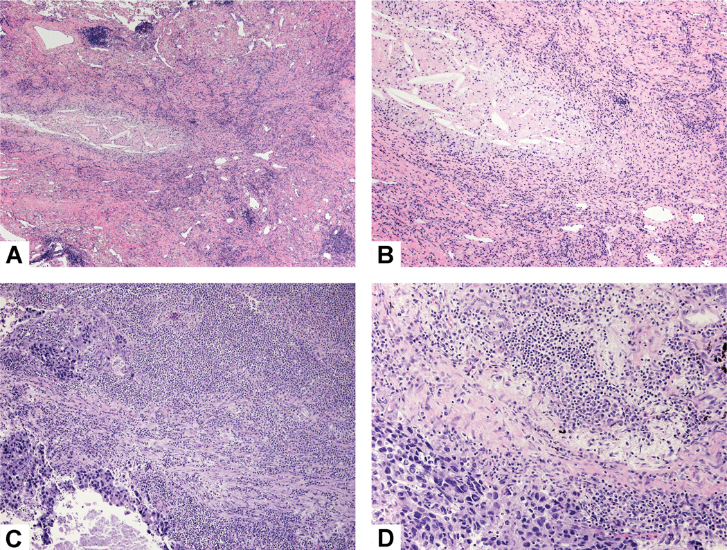
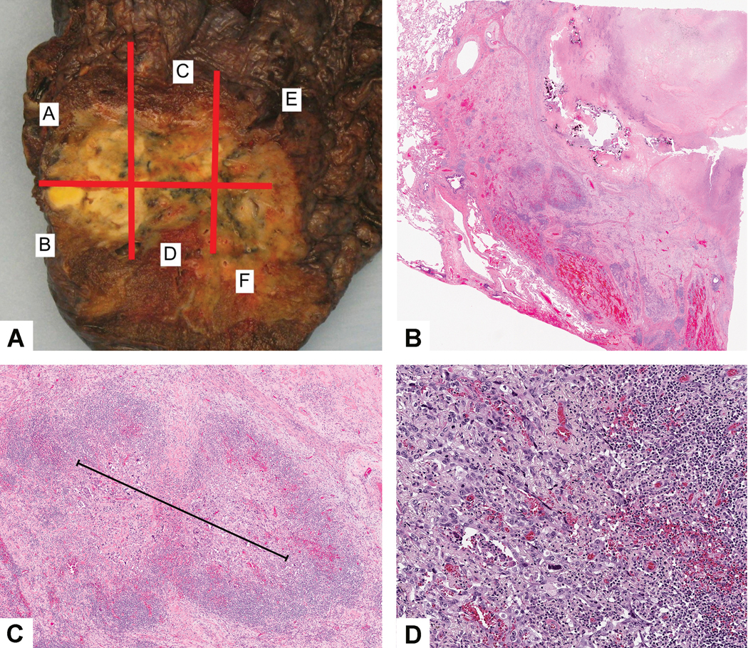
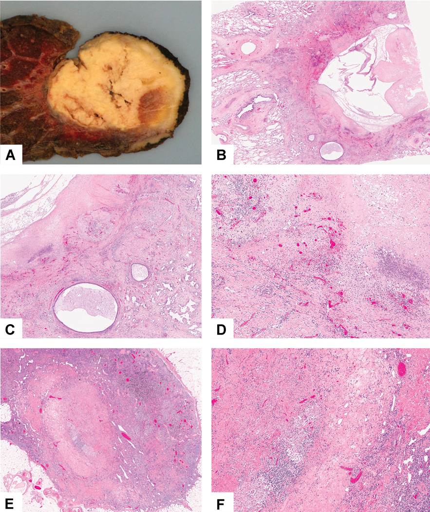
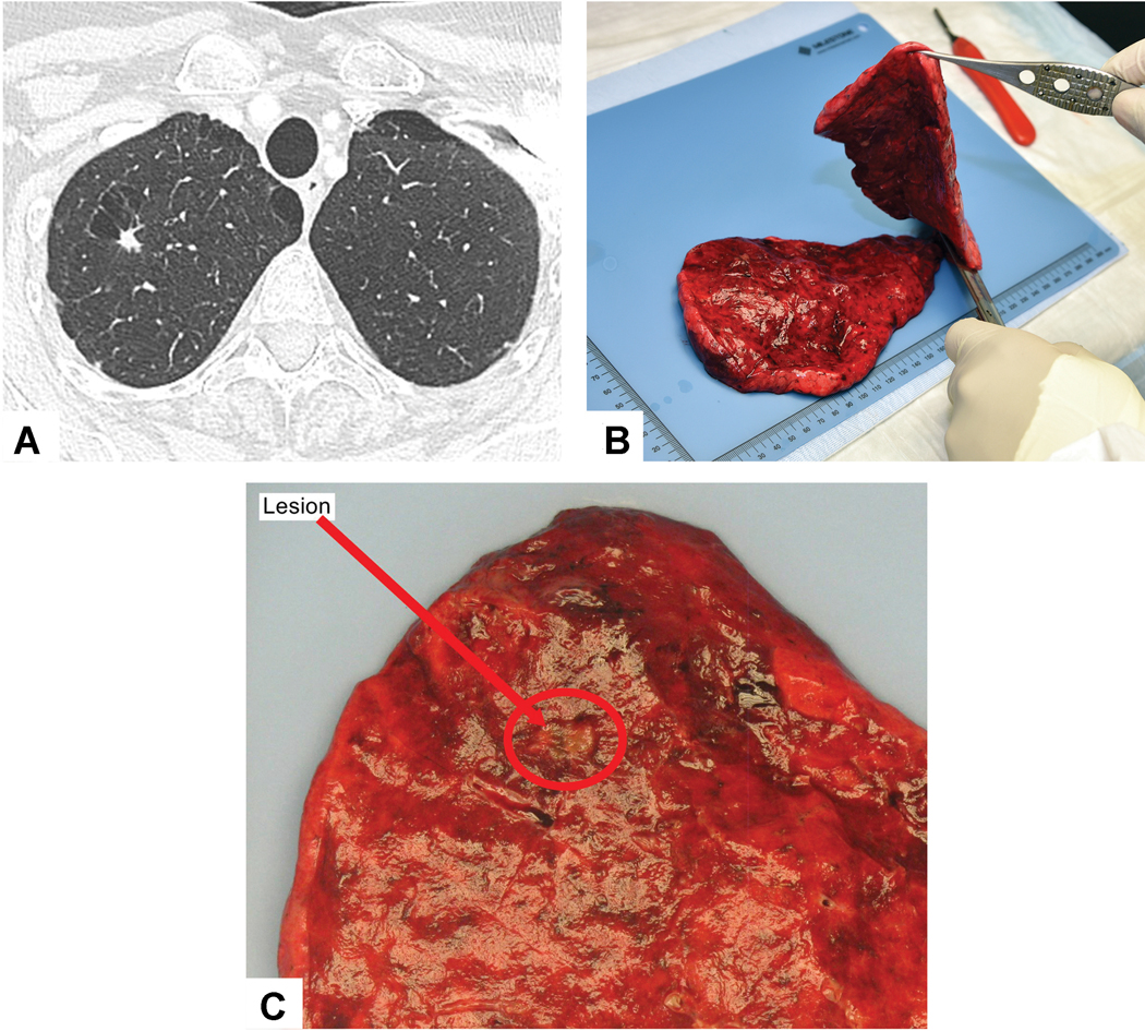
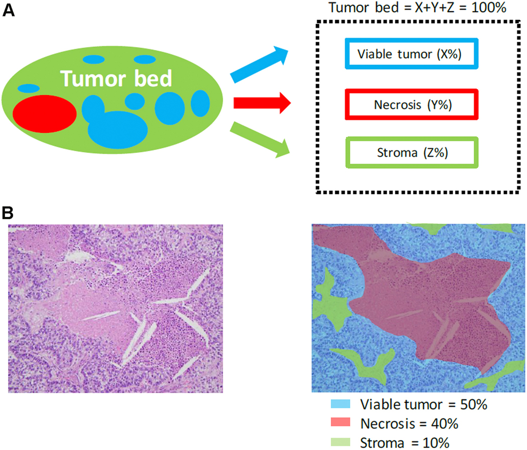
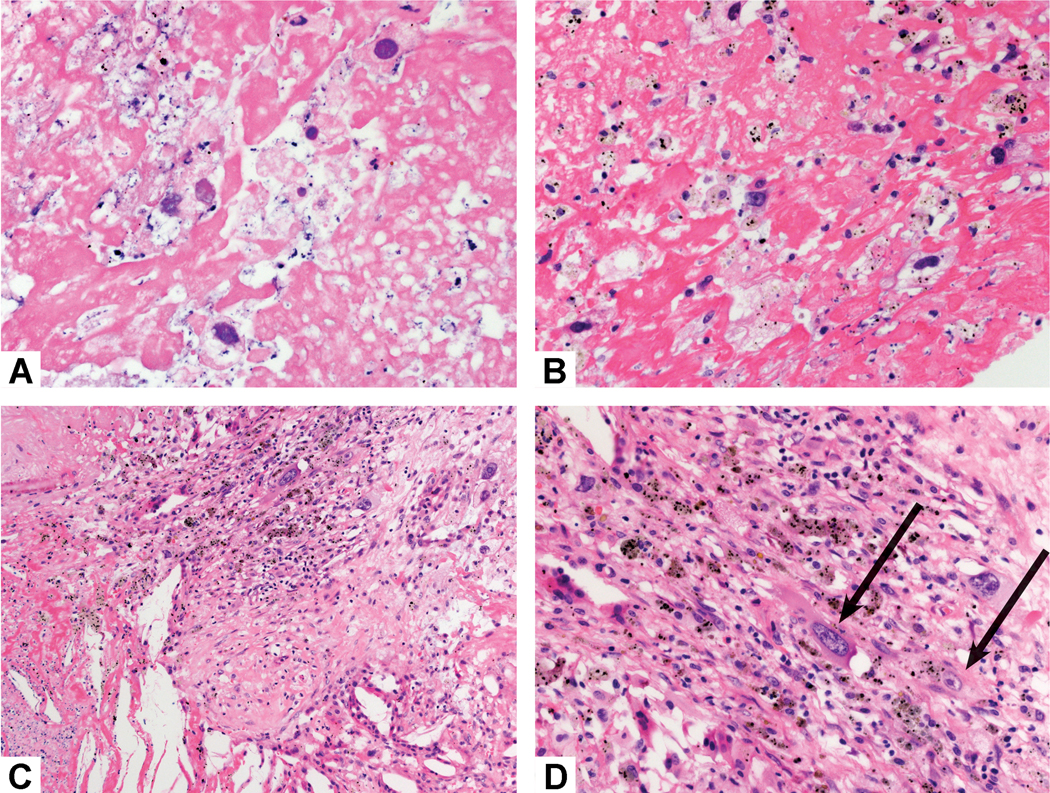
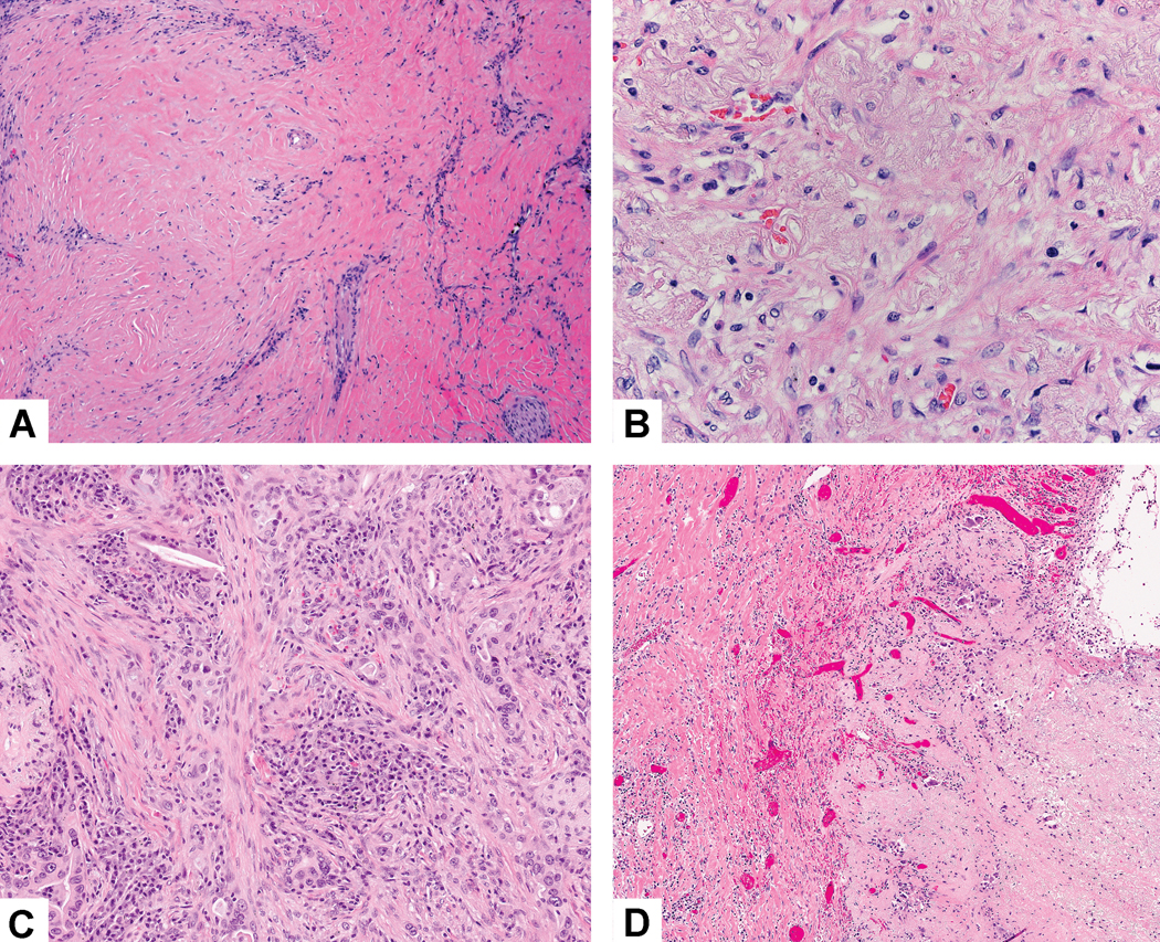
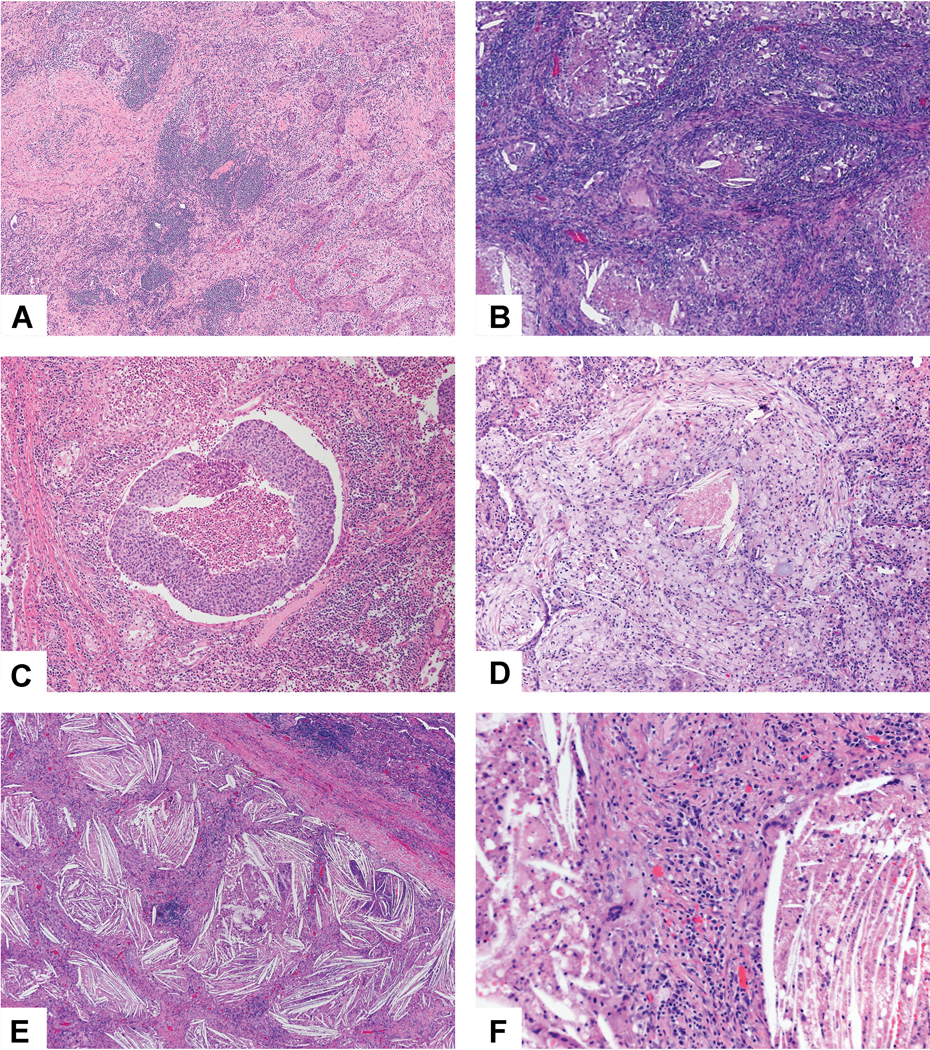
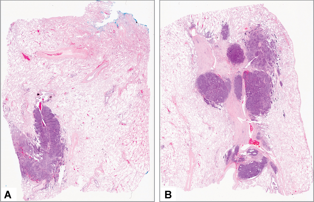
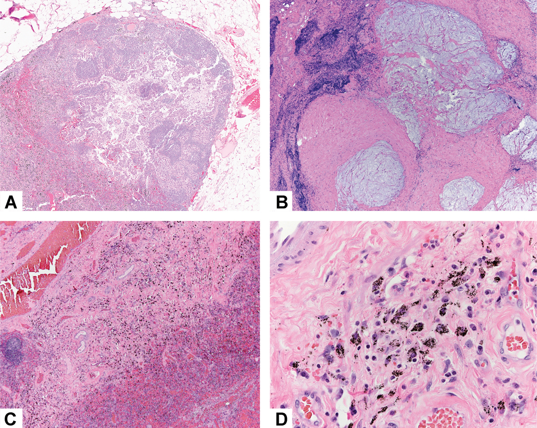
Currently, there is no established guidance on how to process and evaluate resected lung cancer specimens after neoadjuvant therapy in the setting of clinical trials and clinical practice. There is also a lack of precise definitions on the degree of pathologic response, including major pathologic response or complete pathologic response. For other cancers such as osteosarcoma and colorectal, breast, and esophageal carcinomas, there have been multiple studies investigating pathologic assessment of the effects of neoadjuvant therapy, including some detailed recommendations on how to handle these specimens. A comprehensive mapping approach to gross and histologic processing of osteosarcomas after induction therapy has been used for over 40 years. The purpose of this article is to outline detailed recommendations on how to process lung cancer resection specimens and to define pathologic response, including major pathologic response or complete pathologic response after neoadjuvant therapy. A standardized approach is recommended to assess the percentages of (1) viable tumor, (2) necrosis, and (3) stroma (including inflammation and fibrosis) with a total adding up to 100%. This is recommended for all systemic therapies, including chemotherapy, chemoradiation, molecular-targeted therapy, immunotherapy, or any future novel therapies yet to be discovered, whether administered alone or in combination. Specific issues may differ for certain therapies such as immunotherapy, but the grossing process should be similar, and the histologic evaluation should contain these basic elements. Standard pathologic response assessment should allow for comparisons between different therapies and correlations with disease-free survival and overall survival in ongoing and future trials. The International Association for the Study of Lung Cancer has an effort to collect such data from existing and future clinical trials. These recommendations are intended as guidance for clinical trials, although it is hoped they can be viewed as suggestion for good clinical practice outside of clinical trials, to improve consistency of pathologic assessment of treatment response.

Keywords: Lung Cancer; Neoadjuvant therapy; Pathology; Resection specimens; Specimen processing; Treatment response.


Figures

Figure 1:
Figure 1:
Histologic changes without neoadjuvant therapy: A) Only 10% of this tumor was viable with 90% showing fibrosis, chronic inflammation and focal necrosis with cholesterol clefts. B) Higher power shows dense fibrosis with a mild chronic inflammatory infiltrate and an area of necrosis with cholesterol clefts. C) This lung cancer was associated with a large chronic inflammatory infiltrate that overshadowed a small focus of adenocarcinoma. D) This area of solid nests of tumor cells (bottom left) are surrounded by an extensive mixed inflammatory infiltrate of lymphocytes, plasma cells and histiocytes.
Figure 2:
Figure 2:
Major pathologic response: A) This tumor shows a variegated cut surface with yellow and white necrotic areas. B) Low power shows a large area of necrosis surrounded by dense fibrosis and chronic inflammation. C) Only a single 2 mm focus of viable adenocarcinoma was seen. Black line indicates the tumor size measurement. D) This focus of viable tumor cells surrounded by stroma with marked chronic inflammation consists of solid adenocarcinoma that was TTF-1 positive.
Figure 3:
Figure 3:
Complete pathologic response: A) This tumor is almost entirely necrotic with yellow soft areas and small firm tan areas. B) Low power shows a large necrotic cavitated area surrounded by dense fibrosis. C) The stroma shows marked chronic inflammation, foamy histiocytes and fibrosis with some adjacent alveolar parenchyma showing reactive pneumocytes (bottom) D) High power shows numerous chronic inflammatory cells, foamy histiocytes and dense fibrosis. No viable tumor was seen. E) In addition, no tumor was seen in two lymph nodes with changes consistent with treatment effect. This lymph node shows nodular scar and granulomatous inflammation. F) The tumor bed shows dense fibrosis with focal chronic inflammation and giant cells.
Figure 4:
Figure 4:
Gross Assessment to identify the tumor bed: A) Review of the CT scan shows a small 0.8 cm right upper lobe mass. B) After the tumor was identified on palpation, the specimen is cut with a knife through the tumor bed along its maximal dimension. C) This tumor bed measured 0.8 cm and corresponded well to the nodule seen on CT (Red circle highlights the tumor bed).
Figure 5:
Figure 5:
Gross Appearance and Mapping. A) This adenocarcinoma shows extensive necrosis with both yellow areas and some chalky white tissue within areas of cavitation. In addition, this photograph demonstrates the anatomic relationship of the tumor to the overlying pleura and the proximal bronchus. B) This large necrotic tumor shows a large central area of cavitation. In the cavitated area it is impossible to submit blocks of tissue sections, so this area is not mapped. C) This small tumor is mapped but due to its small size the entire tumor was sampled histologically. D) In this case the mapped area turned out to be granulomatous inflammation, and the tumor was on the opposite side of the specimen (red oval shaped circle). So additional sections from the non-mapped area of tumor needed to be submitted to evaluate for treatment effect.
Figure 6:
Figure 6:
Tumor bed with multiple areas of viable tumor. The viable tumor alternated with stromal inflammation and fibrosis that led the person grossing the specimen to describe three different areas of tumor (in blocks 9–4, 9–5 and 9–7) raising the question whether there were intrapulmonary metastases. An area in block 9–6 shows grossly positive lymph nodes reflecting metastatic carcinoma. B) Residual viable tumor consists of acinar glands and the adjacent stroma shows prominent chronic inflammation and loose myxoid connective tissue. C) The nodules of residual viable tumor alternated with intervening fibroinflammatory stroma in the tumor bed. D) Prechemotherapy CT scan shows a solitary mass confirming that there is a single tumor with a heterogeneous response to chemotherapy.
Figure 7:
Figure 7:
Border of tumor with adjacent non-neoplastic lung: A) This tumor shows a thick rim of reactive change beyond the edge of the tumor bed. The black line demarcates the edge of the tumor bed to the left and the nonneoplastic lung to the right. B) In the rim of reactive lung parenchyma between the tumor border and the surrounding normal lung tissue, there is marked interstitial inflammation, fibrosis and prominent reactive pneumocyte proliferation, however, the overall alveolar architecture is preserved. C) This area shows foamy macrophages within an air space consistent with post obstructive endogenous lipoid pneumonia and prominent hyperplastic pneumocytes. The nodular areas of hyperplastic pneumocytes are situated in alveolar spaces altered by the intervening interstitial fibrosis indicating these are reactive changes rather than direct tumor involvement. D) Focally the nonneoplastic lung shows a marked interstitial lymphoid infiltrate surrounding reactive and hyperplastic epithelial cells. E) Organizing pneumonia at the margin of the tumor bed and in the adjacent lung consists of polypoid plugs of loose connective tissue within distal air spaces. F) In this post-obstructive pneumonia setting there are prominent lymphoid aggregates within the nonneoplastic alveolar parenchyma adjacent to the tumor.
Figure 8:
Figure 8:
Histologic components of the tumor bed. A) Schematic image showing how percentage compositions are assigned. The tumor bed is divided into viable tumor area, necrosis and stroma. Stroma includes inflammation and fibrosis. B) A representative hematoxylin-and-eosin stained slide image (left) and a corresponding color illustration of the distribution of the components (right). The blue, red and black areas represent viable tumor, necrosis and stroma, respectively. Figure 1 from Qu et al. with permission.
Figure 9:
Figure 9:
Necrotic versus Viable Tumor Cells. A and B) These necrotic tumor cells are not viable. Although there are nuclear ghosts, the tumor cell cytoplasm is not visible. C) Adjacent to this area of necrosis (bottom) there are rare viable tumor cells (top), highlighted in part D wher the tumor cell cytoplasm is more clearly evident.
Figure 10:
Figure 10:
Stromal fibrosis. A) This tumor stroma consists of dense hyalinized fibrosis. B) The fibrosis in this area has prominent elastic fibers forming a fibroelastotic scar. C) The fibrosis in this area consists of loose spindle shaped myofibroblastic cells with little collagen. D) In this area of fibrotic scarring there are numerous small capillary sized blood vessels.
Figure 11:
Figure 11:
Stromal inflammation and necrosis. A) This squamous cell carcinoma has numerous lymphoid aggregates in the stroma. B) This adenocarcinoma shows a dense lymphoplasmacytic stromal infiltrate. C) Numerous neutrophils are seen not only within the focus of tumor necrosis in the center of the image, but also within the surrounding stroma. D) The tumor stroma is infiltrated by numerous histocytes which show a small area of necrosis in the center. E) Prominent cholesterol clefts are seen in this area of the tumor bed. F) The cholesterol clefts are surrounded by bands of stroma with prominent chronic inflammation and giant cells, some of which are associated with the cholesterol clefts.
Figure 12:
Figure 12:
Different amount of tumor on separate slides. These two slides A and B show different amounts of tumor with at least twice as much tumor on the slide in part B compared to part A.
Figure 13:
Figure 13:
Lymph node assessment. A) This lymph node contains a focus of metastatic adenocarcinoma. It is difficult to be certain what is inflammation in the stroma because of the background lymphocytes in the lymph node. However there appears to be approximately 40–50% tumor necrosis and no stromal fibrosis is seen. B) In this lymph node metastatic mucinous adenocarcinoma had only pools of mucin but no viable tumor cells. Therefore, this was regarded as no viable tumor and the lymph nodes for this case were classified as ypN0. C) Changes related to metastatic tumor related treatment effect need to be distinguished from silicoanthracotic changes as seen in this lymph node where there are numerous histiocytes. D) On close inspection the histiocytes are filled with anthracotic pigment and on polarization microscopy silica-like particles are frequently seen.

Comment in

References

    1. Huvos AG, Rosen G, Marcove RC. Primary osteogenic sarcoma: pathologic aspects in 20 patients after treatment with chemotherapy en bloc resection, and prosthetic bone replacement. Arch Pathol Lab Med 1977;101:14–18. - PubMed
    1. Raymond AK, Chawla SP, Carrasco CH, et al. Osteosarcoma chemotherapy effect: a prognostic factor. Seminars in diagnostic pathology 1987;4:212–236. - PubMed
    1. Chui MH, Kandel RA, Wong M, et al. Histopathologic Features of Prognostic Significance in High-Grade Osteosarcoma. Arch Pathol Lab Med 2016;140:1231–1242. - PubMed
    1. Ali HR, Dariush A, Provenzano E, et al. Computational pathology of pre-treatment biopsies identifies lymphocyte density as a predictor of response to neoadjuvant chemotherapy in breast cancer. Breast cancer research : BCR 2016;18:21. - PMC - PubMed
    1. Peintinger F, Sinn B, Hatzis C, et al. Reproducibility of residual cancer burden for prognostic assessment of breast cancer after neoadjuvant chemotherapy. Modern pathology : an official journal of the United States and Canadian Academy of Pathology, Inc 2015;28:913–920. - PMC - PubMed

Publication types