. 2020;10(1):101-112.
doi: 10.1016/j.jcmgh.2020.01.009. Epub 2020 Jan 28.

CD74 Signaling Links Inflammation to Intestinal Epithelial Cell Regeneration and Promotes Mucosal Healing

Affiliations

CD74 Signaling Links Inflammation to Intestinal Epithelial Cell Regeneration and Promotes Mucosal Healing

Laura Farr et al. Cell Mol Gastroenterol Hepatol. 2020.

Abstract

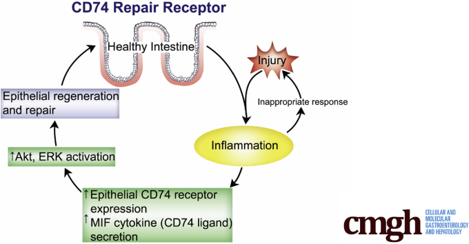
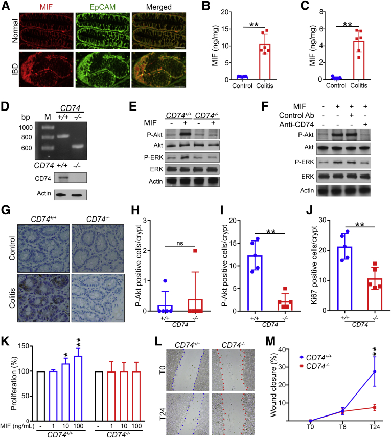
Background & aims: The inflammatory response to intestinal damage promotes healing through mechanisms that are incompletely understood. Gene expression of cluster of differentiation 74 (CD74), the receptor for cytokine macrophage migration inhibitory factor, is increased in patients with inflammatory bowel disease (IBD), however, the role of CD74 signaling in intestinal inflammation remains poorly understood. The aim of this study was to determine the functional role of CD74 signaling in intestinal inflammation.

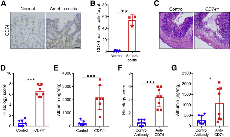
Methods: We studied the characteristics of CD74 protein expression in human IBD and experimental colitis. The functional role of CD74 signaling in the intestine was investigated using cellular models; wild-type, CD74-/-, and bone marrow chimera mice; neutralizing anti-CD74 antibodies; flow cytometry; immunohistochemistry; immunofluorescence; immunoblotting; and clustered regularly interspaced short palindromic repeats and associated protein 9 technology.

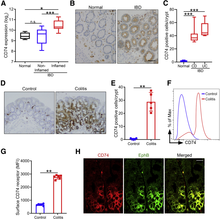
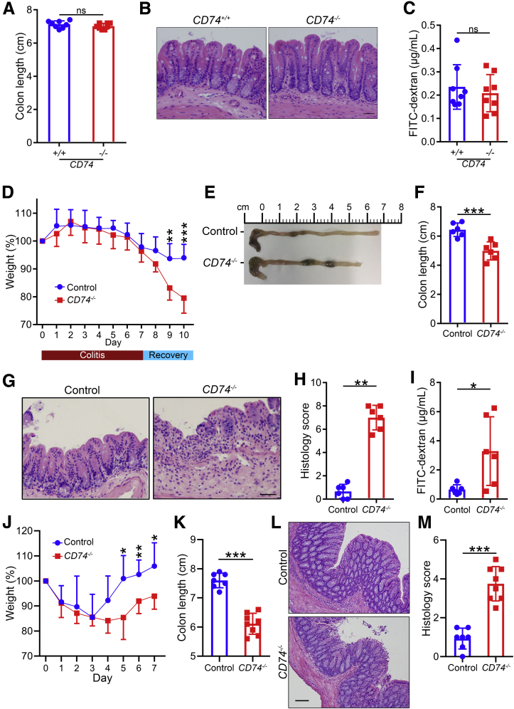
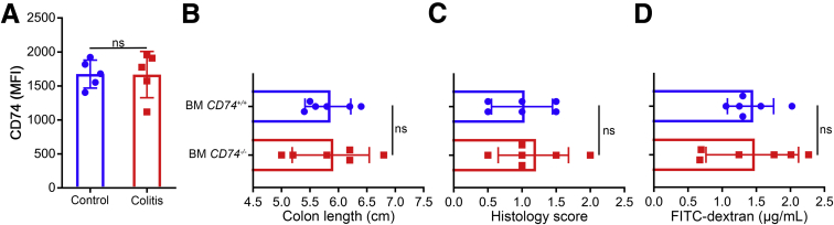
Results: In IBD patients and experimental colitis, CD74-receptor protein expression was increased in inflamed intestinal tissue, prominently in the crypt epithelial cells. By using distinct but complementary chemical and non-chemically induced mouse models of colitis with genetic and antibody neutralization approaches, we found that CD74 signaling was necessary for gut repair. Mechanistically, we found that the macrophage migration inhibitory factor cytokine, which also is increased in colitis, stimulated the CD74 receptor, enhancing intestinal epithelial cell proliferation through activation of the protein kinase B and the extracellular signal-regulated kinase pathways. Our data also suggest that CD74 signaling in immune cells was not essential for mucosal healing.

Conclusions: CD74 signaling is strongly activated during intestinal inflammation and protects the host by promoting epithelial cell regeneration, healing, and maintaining mucosal barrier integrity. Enhancing the CD74 pathway may represent a unique therapeutic strategy for promoting healing in IBD.

Keywords: IBD; MIF Receptor; Proliferation Pathways; Repair.


Figures

None
Graphical abstract
Figure 1
Figure 1
CD74 expression is increased in IBD patients and experimental colitis. (A) Increased CD74 expression in inflamed mucosa. CD74 gene expression levels in normal healthy controls, and in noninflamed and inflamed mucosa of IBD patients (n = 4, 16, and 12, respectively). Bars represent the medians. (B and C) Increased CD74 protein expression in epithelial cells of IBD patients. Immunohistochemical analysis of CD74 expression in mucosa of IBD patients (Crohn’s disease [CD], n = 8; ulcerative colitis [UC], n = 5) and healthy controls (n = 6). Scale bar: 50 μm. (D and E) Increased CD74 protein expression in epithelial cells in experimental colitis. Immunohistochemical analysis of intestinal CD74 expression in mice 24 hours after intracecal injection with 3% DSS in drinking water or normal drinking water control (n = 5 per group). Scale bar: 200 μm. (F and G) Increased surface CD74 expression on intestinal epithelial cells during colitis. Flow cytometry analysis of intestinal epithelial cells for CD74 surface expression (n = 6 per group). (H) Significant CD74 expression in crypt epithelial cells during colitis. Immunofluorescence analysis of intestinal tissue stained with anti-CD74 and EphB antibodies. Scale bar: 20 μm. One representative image of the 3 independent experiments performed is shown. Data represent means and SD. *P < .05, **P < .01, and ***P < .001. MFI, mean fluorescent intensity.
Figure 2
Figure 2
CD74 promotes mucosal healing in DSS- and TNBS-induced colitis. (AC) CD74-deficient mice show normal intestine. Colon lengths, representative H&E-stained images, and fluorescein isothiocyanate (FITC)-dextran levels in WT (CD74+/+) and CD74-deficient mice (CD74–/–) are shown (n = 8 per group). (DI) CD74 promotes repair. Body weight curves, colon lengths, H&E-stained images, and histology scores of WT control and CD74–/– mice treated with DSS followed by a recovery period. Intestinal permeability measured by FITC-dextran in serum 4 hours after gavage (n = 6 per group). (JM) Body weight curves, colon lengths, H&E-stained images, and histology scores of WT control and CD74–/– mice after TNBS-induced chronic colitis (n = 7 and 8, respectively). Scale bars: 100 μm. Data represent means and SD. *P < .05, **P < .01, and ***P < .001.
Figure 3
Figure 3
CD74 promotes mucosal healing in amebic colitis. (A and B) Increased CD74 protein expression in epithelial cells in human amebic colitis. Immunohistochemical analysis of CD74 expression in mucosa of patients with amebic colitis and normal healthy controls (n = 3 and 6, respectively). (CE) H&E-stained images and histology scores of WT control and CD74–/– mice after infection with E histolytica. Intestinal permeability measured by albumin levels in cecal luminal contents (n = 7 per group). (F and G) H&E-stained images, histology scores, and albumin levels in WT mice treated with anti-CD74 or control antibodies (n = 8 per group). Scale bars: 200 μm. Data represent means and SD. *P < .05, **P < .01, ***P < .001.
Figure 4
Figure 4
CD74 signaling activates the pro-proliferative pathways. (A) Confocal immunofluorescence images of intestinal epithelial cells of IBD patient and normal healthy controls expressing MIF. Scale bars: 50 μm. (B and C) Increased MIF secretion in colitis. Intestinal tissue and luminal MIF levels in mice 24 hours after intracecal injection with 3% DSS in drinking water or normal drinking water control (n = 6 per group). (D) Genotyping and immunoblot analysis of CD74+/+ and CD74–/– intestinal epithelial cells. (E and F) CD74-MIF interaction activates Akt and ERK pathways. Immunoblot analysis of Akt and ERK phosphorylation in CD74+/+ and CD74–/– intestinal epithelial cells stimulated with MIF. Akt and ERK phosphorylation assessed by immunoblot analysis of intestinal epithelial cells stimulated with MIF in the presence of anti-CD74 or control antibodies. Actin served as a loading control. (G) Immunohistochemical analysis of Akt phosphorylation in epithelial cells of CD74+/+ and CD74–/– mice during (H) steady-state control and (I) colitis conditions (n = 5 per group). (J) Intestinal tissue stained for Ki67 by immunohistochemistry and quantitated. Scale bars: 50 μm. (KM) CD74 stimulation promotes epithelial cell proliferation and wound closure. Percentage proliferation of CD74+/+ and CD74–/– intestinal epithelial cells 24 hours after dose-dependent stimulation with MIF. Representative images and quantitation of CD74+/+ and CD74–/– intestinal epithelial cell wound closure at baseline (T0) and after 6 hours (T6) and 24 hours (T24) of MIF (100 ng/mL) stimulation. Data are representative of at least 2 independent experiments. Data represent means and SD. *P < .05,**P < .01. Ab, antibody; EpCAM, epithelial cell adhesion molecule; P-Akt, phosphorylated Akt; P-ERK, phosphorylated ERK.
Figure 5
Figure 5
Role of CD74 signaling in immune cells. (A) Flow cytometry analysis of immune cells for CD74 surface expression after 3% DSS in drinking water or normal drinking water control (n = 5 per group). (B) Colon lengths, (C) histology scores, and (D) fluorescein isothiocyanate (FITC)-dextran levels of mice with CD74+/+ and CD74–/– bone marrow (BM) treated with DSS followed by a recovery period (n = 6 per group). Data represent means and SD. MFI, mean fluorescent intensity.

Comment in

  • Here to Heal: Mucosal CD74 Signaling in Colitis.
    Hoffman JM, Rankin CR, Pothoulakis C. Hoffman JM, et al. Cell Mol Gastroenterol Hepatol. 2020;10(1):197-198. doi: 10.1016/j.jcmgh.2020.03.002. Epub 2020 Mar 24. Cell Mol Gastroenterol Hepatol. 2020. PMID: 32220559 Free PMC article. No abstract available.

References

    1. Ng S.C., Shi H.Y., Hamidi N., Underwood F.E., Tang W., Benchimol E.I., Panaccione R., Ghosh S., Wu J.C., Chan F.K. Worldwide incidence and prevalence of inflammatory bowel disease in the 21st century: a systematic review of population-based studies. Lancet. 2017;390:2769–2778. - PubMed
    1. Molodecky N.A., Soon S., Rabi D.M., Ghali W.A., Ferris M., Chernoff G., Benchimol E.I., Panaccione R., Ghosh S., Barkema H.W. Increasing incidence and prevalence of the inflammatory bowel diseases with time, based on systematic review. Gastroenterology. 2012;142:46–54. e42. - PubMed
    1. Nielsen O.H., Ainsworth M.A. Tumor necrosis factor inhibitors for inflammatory bowel disease. N Engl J Med. 2013;369:754–762. - PubMed
    1. Neurath M.F. Current and emerging therapeutic targets for IBD. Nate Rev Gastroenterol Hepatol. 2017;14:269. - PubMed
    1. Neurath M.F., Travis S.P. Mucosal healing in inflammatory bowel diseases: a systematic review. Gut. 2012;61:1619–1635. - PubMed

Publication types

MeSH terms

Substances