. 2020 Jan 31;20(1):85.
doi: 10.1186/s12885-020-6574-4.

Prediction of blood-based biomarkers and subsequent design of bisulfite PCR-LDR-qPCR assay for breast cancer detection

Affiliations

Prediction of blood-based biomarkers and subsequent design of bisulfite PCR-LDR-qPCR assay for breast cancer detection

Manny D Bacolod et al. BMC Cancer. .

Abstract

Background: Interrogation of site-specific CpG methylation in circulating tumor DNAs (ctDNAs) has been employed in a number of studies for early detection of breast cancer (BrCa). In many of these studies, the markers were identified based on known biology of BrCa progression, and interrogated using methyl-specific PCR (MSP), a technique involving bisulfite conversion, PCR, and qPCR.

Methods: In this report, we are demonstrating the development of a novel assay (Multiplex Bisulfite PCR-LDR-qPCR) which can potentially offer improvements to MSP, by integrating additional steps such as ligase detection reaction (LDR), methylated CpG target enrichment, carryover protection (use of uracil DNA glycosylase), and minimization of primer-dimer formation (use of ribose primers and RNAseH2). The assay is designed to for breast cancer-specific CpG markers identified through integrated analyses of publicly available genome-wide methylation datasets for 31 types of primary tumors (including BrCa), as well as matching normal tissues, and peripheral blood.

Results: Our results indicate that the PCR-LDR-qPCR assay is capable of detecting ~ 30 methylated copies of each of 3 BrCa-specific CpG markers, when mixed with excess amount unmethylated CpG markers (~ 3000 copies each), which is a reasonable approximation of BrCa ctDNA overwhelmed with peripheral blood cell-free DNA (cfDNA) when isolated from patient plasma. The bioinformatically-identified CpG markers are located in promoter regions of NR5A2 and PRKCB, and a non-coding region of chromosome 1 (upstream of EFNA3). Additional bioinformatic analyses would reveal that these methylation markers are independent of patient race and age, and positively associated with signaling pathways associated with BrCa progression (such as those related to retinoid nuclear receptor, PTEN, p53, pRB, and p27).

Conclusion: This report demonstrates the potential utilization of bisulfite PCR-LDR-qPCR assay, along with bioinformatically-driven biomarker discovery, in blood-based BrCa detection.

Keywords: Biomarker; Breast cancer; Early detection; Ligase detection reaction; Methylation.


Conflict of interest statement

MDB, AHM, JH, PBF, SFG and FB are shareholders in AcuamarkDx.

Figures
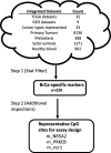
Fig. 1
Fig. 1
The scheme employed to identify potential site-specific methylation markers for blood-based early detection of breast cancer
Fig. 2
Fig. 2
Comparative methylation levels (β values) of the 3 CpG sites (interrogated in the multiplex assay) in breast (BRCA) and other major cancer types among women: colorectal (COADREAD), ovarian (OV), endometrial (UCEC), lung (LUAD, LUSC), and pancreatic (PAAD). The β values range from − 0.5 (0%methyaltion) to + 0.5 (100% methylation)
Fig. 3
Fig. 3
Heatmaps depicting the genes whose transcript levels in breast cancer samples are most highly correlated (negative or positive) with the methylation at the 3 select CpG sites: a) m_PRKCB, b) m_NR5A2, c) m_ncr1
Fig. 4
Fig. 4
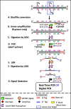
Schematic of the Bisulfite PCR-LDR-qPCR assay for detection of CpG methylation
Fig. 5
Fig. 5
a. Panels 1 and 2 refer to the resulting Ct plots (from ViiA7 run) for multiplex detection of the CpG markers m_ncr1, m_NR5A2, and m_PRKCB using as initial template fragmented and bisulfite-converted mixture of 30, and 3000 genomic copies of DNA from breast cancer cell line (MCF7 or MDA-MB-134-VI) and normal human blood (Roche human genomic DNA) respectively. The DNA fragment mixture simulates the likely constitution of patient cfDNA (i.e. majority of which are released by peripheral blood cells). Panel 3 serves as a negative control (3000 copies of genomic DNA from normal human blood). b. The Ct values for the plots depicted in A. Also shown are results from no template controls (NTCs) in various steps of the assay (PCR, LDR, qPCR). “No Ct” means no amplification was detected after 45 cycles of real time PCR. c. The fraction of methylation at a specific CpG site for the 3 CpG sites in the genomes of MCF7 and MDA-MB-134-VI cell lines, as extracted from Illumina 450 K array-generated datasets deposited in Gene Expression Omnibus (GEO). * Average values extracted from datasets GSE57342, GSE68379, GSE78875, and GSE94943. **Value extracted from dataset GSE68379
Fig. 6
Fig. 6
a. Panels 1 and 2 refer to the resulting Ct plots (from ViiA7 run) for multiplex detection of the CpG marker m_GRK7, using as template 30 copies of bisulfite-converted and fragmented genomic DNA from breast cancer cell line (MCF7 or MDA-MB-134-VI) mixed with 3000 copies of genomic DNA from human blood (Roche human genomic DNA). Panel 3 refers to negative control, with just the normal blood genomic DNA as a template. b. A digital PCR readout (using Formulatrix Constellation dPCR System) for similar experiments depicted in A. NTC refers to “No template control”. c. The fraction of methylation at a specific CpG site for m_GRK7 CpG site in the genomes of MCF7 and MDA-MB-134-VI cell lines. This information was extracted from Illumina 450 K array-generated datasets deposited in Gene Expression Omnibus (GEO). * Average value extracted from datasets GSE57342, GSE68379, GSE78875, and GSE94943. **Value extracted from dataset GSE68379

Similar articles

Cited by

References

    1. Siegel RL, Miller KD, Jemal A. Cancer statistics, 2019. CA Cancer J Clin. 2019;69(1):7–34. doi: 10.3322/caac.21551. - DOI - PubMed
    1. Bray F, Ferlay J, Soerjomataram I, Siegel RL, Torre LA, Jemal A. Global cancer statistics 2018: GLOBOCAN estimates of incidence and mortality worldwide for 36 cancers in 185 countries. CA Cancer J Clin. 2018;68(6):394–424. doi: 10.3322/caac.21492. - DOI - PubMed
    1. Tang Q, Cheng J, Cao X, Surowy H, Burwinkel B. Blood-based DNA methylation as biomarker for breast cancer: a systematic review. Clin Epigenetics. 2016;8:115. doi: 10.1186/s13148-016-0282-6. - DOI - PMC - PubMed
    1. Antoniou A, Pharoah PD, Narod S, Risch HA, Eyfjord JE, Hopper JL, Loman N, Olsson H, Johannsson O, Borg A, et al. Average risks of breast and ovarian cancer associated with BRCA1 or BRCA2 mutations detected in case series unselected for family history: a combined analysis of 22 studies. Am J Hum Genet. 2003;72(5):1117–1130. doi: 10.1086/375033. - DOI - PMC - PubMed
    1. O'Shaughnessy J. Extending survival with chemotherapy in metastatic breast cancer. Oncologist. 2005;10(Suppl 3):20–29. doi: 10.1634/theoncologist.10-90003-20. - DOI - PubMed